Fraud Blocker

Frequently Asked Questions

Frequently Asked Questions

REGISTRATION AND COST QUESTIONS

How do I sign up for a course?

Signing up for any of our LIVE or ONLINE programs is easy!  Once you find your Workshop or Online Package, add it to your cart and complete your purchase!  It’s that easy.

  • You can register for any live course HERE.

No problem!  Your employer can easily register for your – we just have a few tips:  1) The credit card they use MUST match their name and address (including zip code and phone number).  2) They should include your name and email address in the “Additional Information section.”

Registration by check is very easy!  Contact us at (321) 506-9303 and we will send you a form to mail in your payment.

Information for our 2-Day Skills and Procedure Workshop can be found be downloaded by clicking the link below:

Emergency Medicine and Urgent Care Development, Board Review Series, Exam Prep, Question Bank

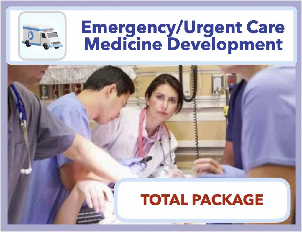
Information for our Emergency Medicine/Urgent Care Development Package can be found by clicking the link below:

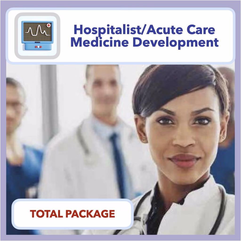
Information for our Hospital Medicine/Acute Care Development Package can be found by clicking the link below:

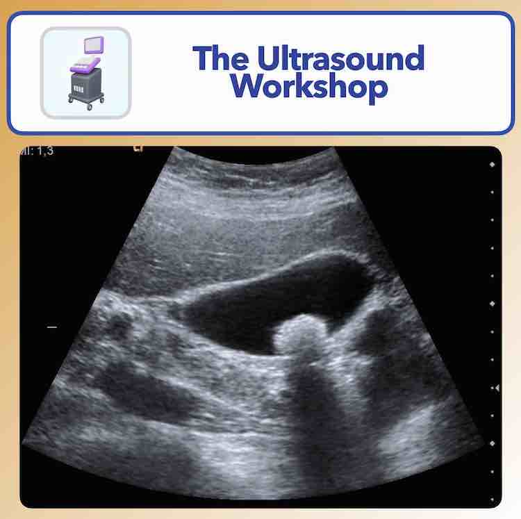
Information for our 1-Day Ultrasound  Workshop can be found be downloaded by clicking the link below:

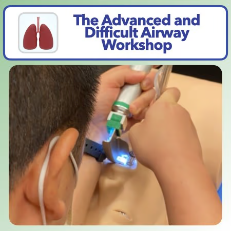
Information for our 1-Day Airway Workshop can be found be downloaded by clicking the link below:

Information for our 1-Day Suturing  Workshop can be found be downloaded by clicking the link below:

  • Each live program ranges from $995 to $2595.00 and includes either a 1-day course, 2-day Clinical Skills and Procedure Workshop, or a 2 or 3-day combination of both programs.
  • All of live programs include a hands on procedure lab, 1 year online access to our website and clinical content, additional CME credits, optional online certification, and a letter of reference.
  • The 2-day clinical skills and procedure course also includes a practice suture kit, tote bag, and optional course companion and reference textbook.
  • Yes – we offer regular promotions and discounts.  These can be found on our promo page.
  • Discounted pricing is posted in real time on our website.
  • Group discounts are offered for groups of 3 or more who book at the same time, and range from 15-25% based on the size of the group.
  • You can also contact us directly and ask us nicely 🙂
  • We do not include hotel costs in our programs, but discounted room rates are available and can be booked directly through our website at the time of registration.
  • You may stay anywhere you choose for our program.
  • You will be responsible for coordinating your own travel arrangements.
  • The POCKET course companion is a printed booklet with a collection of reference normal findings (EKG and radiographs) that you can use during the program to compare abnormal findings shown to the normal studies that you need to compare.  It can also be used during your clinical practice!  This is optional and can be purchased at the time of your registration.
  • A comprehensive course book is included with the live 2-day program and the ultrasound course, and includes all program slides in high quality color, and space for taking notes.
  • All of our programs also have recorded lectures included in the Toolkit, which can reviewed at any time for 1 year.

Our terms and Conditions can be found HERE.

Our Privacy Policy can be found HERE

Rescheduling Policy

The Customer understands that by reserving a seat in one of the company’s programs, they are assuming liability for changes that may effect course income, either directly or indirectly.  The Customer further understands that the costs incurred by the corporation to hold a single event may include contracted food and beverage minimum fees, minimum room rental blocks, and other expenses that are budgeted, in advance, before the course date – often times months in advance.  The Customer understands that rescheduling of their reservation (makeup course) may subject the corporation to lost deposits, fees, and penalties charged by hotels and/or venues, which may not be recoverable at certain venues.  For these reasons, ALL REQUESTS TO RESCHEDULE ATTENDANCE FROM A PREVIOUSLY RESERVED EVENT WILL REQUIRE A $250.00 RESCHEDULING FEE.  THIS INCLUDES ADDITIONAL EVENTS THAT HAVE BEEN PREVIOUSLY RESCHEDULED AND ARE REQUESTED TO BE RESCHEDULED AGAIN.  In addition, PREPAYMENTS FOR HOTEL COSTS MADE TO THE COMPANY WILL BE NON-REFUNDABLE.  The Customer understands that a repeat payment for hotel fees will be required if overnight lodging is requested, and will be determined by the company’s advertised rate at the time of registration. The Corporation reserves the right to reschedule courses based on unexpected conditions, natural disasters, disease outbreaks, epidemics, pandemics, or other unforeseeable events that are detrimental to the operational safety of the scheduled program.  In  the event such cancellation is necessary, Customers will be rescheduled for the next available program at their convenience.  A maximum of 1 scheduled change is permitted per customer unless approved by the Corporation.  All rescheduling changes must occur within one calendar year from the original date reserved and may not be permitted based on occupancy of the replacement date requested.  This will be at the sole discretion of the Company.  The Customer understands and agrees that ALL FEES PAID TO ATTEND A PROGRAM MAY BE NON-REFUNDABLE IF THEY ARE UNABLE TO ATTEND THE PROGRAM FOR WHICH THEY ARE REGISTERED.

Refund Policy

All requests for refunds must be made in writing or email format.  Refunds for program fees (defined as the total sum of any payments made excluding hotel reservations) are determined based on a pro-rated schedule prior to the date of the program as follows:

91 or more days prior to the program date:  All program fees are fully refundable minus a $250 reservation fee.

90 to 61 days prior to the program date:  50% of the total program fees will be eligible for a refund 

60 to 31 days prior to the program date:  25% of the total program fees will be eligible for a refund  

NO REFUNDS WILL BE GIVEN FOR CUSTOMER CANCELLATIONS WITHIN 30 DAYS OF THE SCHEDULED EVENT.  

HOTEL BOOKING FEES ARE NOT ELIGIBLE FOR REFUNDS REGARDLESS OF DATE OF REFUND REQUEST.

ALL ONLINE CONTENT PURCHASES (INCLUDING MEMBERSHIPS, REVIEW PROGRAMS, LECTURES, OR OTHER ITEMS PURCHASED THROUGH THE COMPANY WEBSITE OR ITS REFERRAL SOURCES, AND NOT DESIGNATED AS LIVE EVENTS) ARE NON-REFUNDABLE.  ALL SALES ARE FINAL FOR THESE PRODUCTS.

CONTENT AND SCOPE QUESTIONS

Should I take this course?

Yes!  This program is designed for Nurse Practitioners, Physician Associates, Physicians, Residents, and Students.  We teach advanced and complex concepts that anyone practicing primary care, urgent care, emergency, hospitalist, acute care, or any hospital-based medicine should know!

We guarantee you will walk away with new concepts, new ways of looking at things you haven’t understood, and the confidence your patients deserve!

  • Participants in this course will have a clear understanding of how to identify common emergencies, perform primary care procedures, perform emergency procedures and life-saving interventions, and apply these in ANY PRACTICE SETTING.

Yes!

  • Yes – all of our programs are accredited for CME credits that are interchangeable with most nursing and physician assistant certifying organizations.
  • All program purchases include membership to our online Clinical Toolkit, which has additional accredited CME opportunities for you.
  • Content is added on a regular basis.
  • Physicians may claim credit from the American Academy of Family Physicians, which count toward the Physician Recognition Award (PRA) as Category I AMA credits.
The table below breaks down all CME credits, by product:
Category I CME Credits Emergency Procedure Credits Pharmacology Credits
1-Day Airway Course (Live or Online) 10 8.5 1.5
1 -Day Ultrasound Course (Live or Online) 14 10 1.5
1-Day Advanced Suturing Workshop 12 10 1
2-Day Clinical Skills and Procedure Workshop 23 12 3
3-Day Airway, Skills, and Procedure Workshop 33 20.5 4.5
3-Day Ultrasound, Skills, and Procedure Workshop 37 22 4.5
4-Day Virtual Conference 47 30.5 6
Emergency Medicine QBank 30 10 18
Emergency Medicine ENP/CAQ Core Review Series 60 10 7
Hospitalist Medicine Qbank 40 0 18
Hospitalist Medicine CAQ Core Review Series* 40 0 7
Online ECG and Cardiology Course 25 10 15
Inpatient Guide 12 10 1
*Also includes risk management module.

This activity has been reviewed by the American Academy of Physician Associates Review Panel and is compliant with AAPA CME Criteria. This activity is designated for 324 AAPA Category 1 CME credits. PAs should only claim credit commensurate with the extent of their participation. Approval is valid from 2/20/2025 to 2/20/2026. AAPA reference number: CME-2013021.

The AAFP has reviewed the Physician Skills and Procedures Series, and deemed it acceptable for AAFP credit. Term of approval is from 05/01/2025 to 04/30/2026. Physicians should claim only the credit commensurate with the extent of their participation in the activity.

From the AAPA Website:

NPs can often claim AAPA Category 1 CME credit as continuing education with AANPCB and ANCC. AAPA recommends that NPs check with their certification/licensing authority to confirm that AAPA credit is acceptable.

From the AAFP Website:

AAFP Prescribed credit is accepted by the following organizations. Please contact them directly about how participants should report the credit they earned.

  • American Academy of Physician Assistants (AAPA)
  • National Commission on Certification of Physician Assistants (NCCPA)
  • American Nurses Credentialing Center (ANCC)
  • American Academy of Nurse Practitioners Certification Board (AANPCB)
  • American Association of Medical Assistants (AAMA)
  • American Board of Family Medicine (ABFM)
  • American Board of Emergency Medicine (ABEM)
  • American Board of Preventative Medicine (ABPM)
  • American Board of Urology (ABU)

1-day programs begin at 8am and end around 5pm.  Your specific agenda can be found on the program page where you would register:

Ultrasound Workshop

Airway Workshop

Advanced Suturing Workshop

2-day programs begin at 8am and end around 5pm each day.  Your specific agenda can be found on the program page where you would register:

Clinical Skills and Procedure Workshop

3-day programs begin at 8am and end around 5pm each day.  Your specific agenda can be found on the program page where you would register:

3-Day Ultrasound, Skills, and Procedure Workshop

3-Day Airway, Skills, and Procedure Workshop

All programs are taught by our award-winning faculty who live locally in the city where your program is held.  We do this to ensure a generous 6:1 student to instructor ratio and to maximize individual attention.

Unlike other programs out there, we have designed our entire program around you as an individual learner and a practitioner who is part of a team.  Our small group format encourages you to ask questions and maximize your time with our faculty.  Our unique blend of well-timed didactics and hands on procedures leaves you immersed each day, and you will leave with confidence at the end of your experience.

  • There is truly nothing out there that does what we do the way we do it, in the amount of time that we do it in, and still provides the resources that we do.
  • A team of physicians and Advanced Practice Providers for each course ensures you will receive balanced education.
  • Our online clinical resources are direct, focused, and not full of fluff seen on other online sites
  • Additional CME opportunities will ensure regular annual CME requirements are met every year

MEMBERSHIP QUESTIONS

Does signing up for a membership get me into the two-day live program?

Basic membership in our Clinical Toolkit is included with any live or online purchase, but does not replace actual program registration.  However, membership in our ALL ACCESS plan includes heavily discounts attendance to any 1 live workshop annually.

Our membership levels come in three tiers:

Basic toolkit membership includes over 300 clinical topics with high-yield reference, deep dive content review, videos, audio, and additional CME opportunities.  Members also have access to our members-only commentary and discussion forums.

Enhanced toolkit membership includes everything in the basic membership plan, along with CME credits for all Basic toolkit content.  This adds an additional 100 CME credits. 

Also includes all required NP certification/renewal credits for Pharmacology and Emergency Procedures

All-access membership includes everything in the Basic and Enhanced membership levels, and adds access to our ENTIRE ONLINE LIBRARY of online packages, and deeply discounted registration to 1 live program of your choice annually.  There is no renewal charge for this plan, and as we add content to our platform, you get it at no charge – for life!

Yes!  You can register for any of our membership levels HERE.

Yes.  All Access membership level members get a significant discount on ALL live programs.

Toolkit memberships start at $149.  Learn more HERE.

You can become a member in one of two ways: 1) Sign up for any live or online workshop or package, or 2) buy a membership plan HERE.

PROGRAM LOCATION QUESTIONS

Where is the program held?

Live programs are held in one of three cities:  Orlando, Florida,  Dallas, Texas,  and Washington, DC.  Specific programs are available in different locations throughout the year.  Hotel information is detailed on each program page.

We do not provide a hotel room booking or reservation service.  However, all registrants are provided with a direct contact to our hotel to make their reservations.

All live programs begin at 8am and end by 5pm.  We encourage you to arrive by 7:45em each day to get settled and help us start on time.

Dress comfortably!  We suggest you bring a long sleeve shirt or sweatshirt if you get cold.  Otherwise, dress however you learn the most!

There is nothing you “need” to attend our programs – we have everything you need ready for you when you arrive.  We do suggest you bring snacks throughout the day, and eat a light breakfast before the program begins.

Access to our Clinical Toolkit is available on the date you registered for your program.  Online programs provide instant access to all content.  Live program participants also have immediate access and are encouraged (but not required) to review content prior to the live program.

ACCREDITATION AND CERTIFICATION QUESTIONS

I took the course. How do I get my certificate?

Claiming CME is easy!  To get your certificate, simply log in to the Clinical Toolkit, find your program on the dashboard, and click on the CME icon for your program.  You will need to complete a post-course survey to claim your credits.  Once the survey is complete, you can download your certificate.  Your certificate will be saved in your account profile for retrieval whenever you need it.  You will need to keep your toolkit membership active to use this service.

Our 2-Day Skills and Procedure Workshop, Airway Workshop, and Ultrasound Workshop include an optional certification exam.  This validated test requires a passing score of 80%.  When you pass, you will get a certificate of completion and a link for your employer or credentialing team to verify your new skills!

Unique to the 2-day Skills and Procedure Workshop, all successful certifications will also award a signed letter of reference, which you can use for your job search and to verify that you went above and beyond to prove your skills!

HOW DO I GET A COPY OF MY CME CERTIFICATE?

Once you complete our program, you will need to complete an online survey which will automatically generate your certificate.  This can be found by logging into the Clinical Toolkit.

CONTACT US

We can be reached by email at Support@ppemedical.com or by telephone at (321) 506-9303

The Advanced and Difficult Airway Course

[tribe_events_list view="photo" category="ADAC"]

The Ultrasound Course

[tribe_events_list view="photo" category="POCUS"]

The Clinical Skills & Procedure Workshop

[tribe_events_list view="photo" category="2DAYEVENT" cost="no" limit="10"]

The Clinical Skills & Procedure Workshop + The Airway Course

[tribe_events_list view="photo" category="ADAC3"]

The Clinical Skills & Procedure Workshop + The Ultrasound Course

[tribe_events_list view="photo" category="POCUS3"]

Dermatology Overview

Dermatology Essentials

Definition

Cellulitis: infection of dermis and subcutaneous fat

Impetigo: superficial purulent lesions, esp. on face and extremities. Commonly with bullae and/or golden crust

Erysipelas: raised erythematous lesion with clear borders

Folliculitis: hair follicle inflammation. Superficial and limited to the epidermis.

Furunculosis: hair follicle infection that extend to dermis. Multiple = carbuncle

Necrotizing Infection: Deeper SSTI that involve fascial and/or muscle compartments

Etiology

Microbiology

  • Cellulitis: primarily Staph and Strep, incl. MRSA. In immunocomp./diabetics, GNRs also
    • Other etiologies: cat/dog bite P. moltocida; gardening Sporothrix; salt water Vibrio vulnificus; puncture wound → Pseudomonas
  • Impetigo: Strep or Staph
  • Erysipelas: group A Strep usu.
  • Folliculitis/furunculosis: S. aureus, Pseudomonas
  • Necrotizing Infections: Polymicrobial (eg strep and GNRs in Type I, Fournier’s), Group A Strep, S. aureus, Aeromonas hydrophila, Vibrio vulnificus

At risk: athletic teams, military, prison, MSM, communities with MRSA infxn, Diabetic

High risk for more aggressive infection: splenectomy, immunocompromised

Differential Diagnosis

  • Cellulitis
  • Impetigo
  • Erysipelas
  • Folliculitis
  • Furunculosis
  • Necrotizing fasciitis
  • Myonecrosis
  • Calciphylaxis
  • Cutaneous metastasis from neoplasms (especially adenocarcinoma)
  • Graft-versus-host disease (in appropriate population)
  • Sweet syndrome

Patient History

  • Recent trauma to the affected area?
  • Any recent surgeries (hip replacement is risk factor)?
  • Ask about the presence of HIV, diabetes, liver disease, or kidney disease.
  • History of IV drug abuse or subcutaneous injection.
  • Recurrent Cellulitis: Assess for predisposing conditions such as edema, obesity, eczema, venous stasis, and toe web abnormalities.
  • Recurrent Abscesses: Search for local causes such as pilonidal cyst, HS, or foreign body. Consider 5-day decolonization (intranasal mupirosin, daily chlorhexidine). Consider neutrophil disorder if abscesses began in childhood.

Physical Exam

  • Evaluate affected area for erythema, edema, warmth, and pain on palpation.
  • Look for lymphangiitis (erythematous tracks under the skin marking an inflamed lymphatic system), palpate for lymphadenopathy.
  • Assess for evidence of necrotizing infection: systemic toxicity with high temperature, hypotension, disorientation, lethargy, skin discoloration or bullous lesions, anesthesia, firm skin with wooden-hard induration, pain extending beyond cutaneous erythema, pain out of proportion to exam

Work Up

Note: Diagnosis is largely clinical

Laboratory:

  • CBC with diff, ESR/CRP if concern for osteo, CK if concern for necrotizing infection or pyomyositis.
  • Furuncle/pustule can be aspirated for gram stain and culture.
  • For cellulitis, blood cultures are generally low yield, but should be obtained in patients undergoing chemo, neutropenic patients, and those who suffered animal bites.

Imaging:

  • If concern for osteo, xray; consider MRI
  • If concern for necrotizing infection can look for gas in fascial planes on x-ray or CT, but this is highly insensitive

Triage

More serious presentations of skin and soft tissue infections:

  • Toxic shock syndrome: fever, HA, vomiting, myalgias, pharyngitis, diarrhea, diffuse rash with desquamation. Hypotension and shock.
  • Osteomyelitis: infection of bone due to hematogenous seeding or direct spread from overlying focus.
  • Necrotizing fasciitis: infection and necrosis of superficial fascia, subq fat, and deep fascia. Clues: rapidly spreading cellulitis, systemic toxicity (inc TSS), pain out of proportion to exam, bullae formation, gangrene, crepitus. Surgical and medical emergency.
  • Gas gangrene: Clostridial myonecrosis, a fulminant skeletal muscle infection. C. perfringins usually in the setting of trauma; C. septicum in setting of cancer. Surgical and medical emergency.

Treatment

Purulent (furuncle/carbuncle/abscess):

  • Mild: I & D
  • Moderate: I & D, send for culture and sensitives
    • Empiric treatment: Bactrim 1-2 DS tab BID or Doxycycline 100mg BID
    • Defined treatment: MRSA: Bactrim 1-2 DS tab BID, MSSA: Dicloxacillin 250 Q6H or Cephalexin 500 Q6H or Cefadroxil 500mg po q12.
  • Severe: I & D, send for culture and sensitiivies
    • Empiric treatment: Vancomycin or Daptomycin or Linezolid or Ceftaroline
    • Defined treatment: MRSA: similar to empiric, MSSA: Nafcillin or Cefazolin or Clindamycin (if Susceptible)

Nonpurulent (necrotizing infection/cellulitis/erysipelas):

  • Mild: impetigo: topical mupirocin; oral treatment: Penicillin VK or Cephalosporin (eg Cephalexin 500mg PO Q6H) or Dicloxacillin 500mg PO Q6H or Clindamycin 300mg PO Q8H
  • Moderate: IV therapy: penicillin or Cefriaxone or Cefazolin or Clindamycin 300mg PO Q8H or 600mg IV Q8H
  • Severe: emergency surgical evaluation/debridement to rule out necrotizing process
    • Empiric treatment: Vancomycin PLUS Piperacillin/Tazobactam
    • Defined treatment for necrotizing infections:
      • Strep. pyogenes:Penicillin PLUS Clindamycin
      • Vibrio vulnificus:Doxycycline PLUS Ceftazidime
      • Aeromonas hydrophila:Doxycycline PLUS Ciprofloxacin
      • Polymicrobial: Vancomycin PLUS Piperacillin/Tazobactam

Duration of Therapy: 5-7 Days

Treatment Notes:

Erythema may initially worsen with antibiotics 2/2 local bacterial killing.

– For cellulitis, elevation of the affected extremity is essential to treatment.

– For Staph aureus infections (eg suppurative cellulitis) in 2014 at Hopkins susceptibilities were: TMP-SMX 87-88%, Tetracycline 89-91%, and Clindamycin 46-60%.

– For Beta-hemolytic Strep infections (eg non-suppurative cellulitis) all strains are susceptible to penicillin. At Hopkins there are high rates of resistance to TMP-SMX and tetracyclines and variable rates of resistance to Clindamycin.

– If you are concerned for a necrotizing infection, CONSULT SURGERY. Empiric antibiotic treatment with vancomycin (or linezolid) PLUS zosyn (or carbapenem) should be initiated. Clindamycin can be added to inhibit toxin production.

References

  1. Stevens DL, Bisno AL, Chambers HF, et al. Practice guidelines for the diagnosis and management of skin and soft tissue infections: 2014 update by the Infectious Diseases Society of America. Clin Infect Dis. 2014;59(2):e10-52. [PMID:24973422]
  2. Swartz MN. Clinical practice. Cellulitis. N Engl J Med. 2004;350(9):904-12. [PMID:14985488]

Resources

Heart Failure

Cardiology Essentials

Definition

Syndrome characterized by impaired myocardial performance and progressive maladaptive neurohormonal activation of the cardiovascular system leading to circulatory insufficiency to meet the body’s demands.

Systolic heart failure or heart failure with reduced ejection fraction (HFrEF): Clinical diagnosis of heart failure and an EF of less than 50%.

Diastolic heart failure or heart failure with preserved ejection fraction (HFpEF): Clinical signs and symptoms of heart failure with evidence of normal or preserved EF and evidence of abnormal LV diastolic function by Doppler echocardiography or cardiac catheterization

Right heart failure: Majority of cases are a result of left heart failure, although isolated pulmonary diseases can also cause this syndrome.

Etiology

  • Non-ischemic dilated cardiomyopathy (familial or idiopathic)
  • Hypertrophic cardiomyopathy
  • Restrictive cardiomyopathy
  • Cardiomyopathy as a result of fibroelastosis
  • Mitochondrial disease
  • Left ventricular non-compaction
  • Ischemic cardiomyopathy
  • Stress induced cardiomyopathy
  • Valvular obstruction or insufficiency
  • Hypertensive cardiomyopathy
  • Inflammatory (lymphocytic, eosinophilic, giant cell myocarditis)
  • Infectious (Chagas, Lyme disease, HIV, viral, bacterial, or fungal infections)
  • Endocrine disorders (thyroid disease, adrenal insufficiency, pheochromocytoma, acromegaly)
  • Familial storage disease (hemochromatosis, glycogen storage disease, Hurler syndrome, Anderson-Fabry disease)
  • Amyloidosis
  • Connective tissue disease (SLE, polyarteritis nodosa, scleroderma, myositis, sarcoidosis)
  • Muscular dystrophies
  • Neuromuscular disease (Friedreich ataxia, Noonan disease)
  • Toxins (alcohol, anthracyclines, radiation)
  • Tachyarrhythmia

Pathophysiology

Progressive disorder initiated by a form of myocardial injury either sudden (MI or myocarditis) or chronic insults (familial, metabolic, HTN, valve disease, shunting) that result in maladaptive compensatory mechanisms.

These mechanisms include activation of the sympathetic nervous system and activation of the RAS system which overtime lead to pump dysfunction and circulatory collapse.

Differential Diagnosis

Other entities that may look like acute decompensated heart failure:

  • Acute coronary syndrome
  • Interstitial lung disease
  • Pneumonia
  • ARDS
  • Other sources of volume overload such as CKD/ESRD vs cirrhosis, pulmonary hypertension, PE, cardiac tamponade, constrictive or restrictive pericarditis

Patient History

Ask about the signs and symptoms:

  • Worsening dyspnea at rest or exertion?
  • Fatigue?
  • Orthopnea?
  • PND?
  • Weight gain?
  • Increased edema?
  • Lightheadedness?
  • indigestion?
  • Chest heaviness?
  • Fever?
  • Chest pain?
  • Timing of symptom onset?

Ask about triggers of acute decompensation:

  • dietary indiscretion? foods high in Na like lunch meats, chips, canned foods, fast foods?
  • missed medication doses (diuretic)?
  • are they weighing themself daily? adjusting diuretics?
  • any signs or symptoms that an ischemic event has occurred?
  • do they consume alcohol excessively?

Physical Exam

  • Weight gain (if possible look at previous discharge weights)
  • Elevated jugular venous pulsations (Key!), hepatojugular reflux
  • Orthopnea
  • Pulmonary rales
  • Third and/or fourth heart sound
  • Pedal edema
  • Sacral edema in patients who are mostly in bed

Work Up

Laboratory

  • Renal function panel, liver function panel (CMP): Patients who are volume overloaded due to acute decompensated heart failure often have an acute kidney injury and hepatic congestion.
  • Potassium, calcium (CMP), magnesium. May need to check more frequently (e.g. bid) especially if pt will be diuresed.
  • CBC: Anemia is present in up to 40% of patient with heart failure.
  • Consider pro-BNP if volume exam not helpful; compare to prior.
  • If patient is presenting newly with HF and/or etiology is unclear:
    • troponin and lipid profile, especially if HFrEF the pt may need further work up for ischemic disease
    • TSH
    • in the right patient, consider iron studies (hemochromatosis), serum ceruloplasmin (Wilson’s), trypanosoma cruzi IgG (chagas), blood alcohol level or CDT etc.

Imaging

  • ECG, chest x-ray, echocardiography

Other imaging and diagnostic modalities that can be considered based on the patient’s history:

  • Cardiac MR
  • Nuclear imaging
  • Right heart catheterization
  • Left heart catheterization
  • CT angiogram.
  • Endomyocardial biopsy


Triage

Strongly consider step-down or ICU if evidence of decompensation with hypoperfusion (cold and wet):

Altered mental status, Cold extremities, evidence of organ hypoperfusion: increasing lactate or rising creatine, narrow pulse pressures

Risk Stratification

The American College of Cardiology/American Heart Association (ACC/AHA) Heart Failure Classification is a system used to classify heart failure into four stages based on the severity of symptoms and degree of functional impairment.

The four stages of heart failure in the ACC/AHA classification are:

  1. Stage A: At high risk of developing heart failure due to underlying conditions or risk factors such as hypertension, diabetes, or coronary artery disease.

  2. Stage B: Structural heart disease is present, but there are no symptoms of heart failure. This stage includes patients with a history of myocardial infarction (heart attack) or left ventricular remodeling after a cardiac injury.

  3. Stage C: Structural heart disease is present, and there are symptoms of heart failure such as fatigue, shortness of breath, and decreased exercise tolerance. This stage includes patients with past or current symptoms of heart failure who are responding to treatment.

  4. Stage D: Advanced heart failure that is refractory to standard treatments. This stage includes patients with severe symptoms of heart failure at rest, despite maximal medical therapy. Patients in this stage may require advanced interventions such as heart transplant or mechanical circulatory support.

The ACC/AHA Heart Failure Classification is based on a combination of factors, including clinical symptoms, physical examination findings, imaging studies, and laboratory tests. This classification system is useful for guiding treatment decisions and predicting outcomes in patients with heart failure. It can also help clinicians identify patients at high risk for developing heart failure and initiate preventive interventions to improve outcomes.

The New York Heart Association (NYHA) Functional Classification is a system used to classify heart failure into four stages based on the severity of symptoms and degree of functional impairment. The classification system was developed in 1928 and is still widely used today.

New York Heart Association functional classification

The NYHA Functional Classification is based on the patient’s subjective symptoms and limitations related to physical activity. It is often used in clinical practice to assess the severity of heart failure, guide treatment decisions, and predict outcomes. Patients with more severe symptoms are more likely to have poorer outcomes, and may require more aggressive treatment or consideration of advanced interventions, such as heart transplantation or mechanical circulatory support.

It’s important to note that the NYHA Functional Classification is just one aspect of the overall assessment of heart failure and should be used in conjunction with other clinical and diagnostic findings.

The Seattle Heart Failure Model (SHFM) is a clinical prediction model that provides an estimate of the probability of death and other adverse outcomes in patients with heart failure. It was developed to help clinicians make more informed decisions about treatment and to assist in risk stratification of patients with heart failure. The SHFM incorporates a wide range of patient characteristics, including demographics, clinical symptoms, laboratory values, and medication use, to predict the likelihood of various outcomes, such as mortality, hospitalization, and quality of life. The model is based on data from over 11,000 patients with heart failure and has been validated in several independent cohorts. To use the SHFM, a clinician inputs data on the patient’s age, sex, symptoms, medical history, laboratory values, and medication use into a web-based calculator. The model then generates a personalized estimate of the patient’s probability of death and other outcomes at 1 year and 5 years. The SHFM also provides a range of other information, such as the estimated survival time, probability of hospitalization, and predicted quality of life. The SHFM has been shown to have good accuracy in predicting outcomes in patients with heart failure, and it can be useful in guiding treatment decisions and in risk stratification of patients. However, it is important to note that the SHFM is just one tool among many that can be used in the management of heart failure, and it should be used in conjunction with clinical judgment and other diagnostic and prognostic tools.  

The MAGGIC (Meta-Analysis Global Group in Chronic Heart Failure) risk score is a prognostic model that is used to predict mortality in patients with chronic heart failure. It was developed using a large international database of over 39,000 patients with heart failure from 30 different studies.

The MAGGIC risk score takes into account a range of patient characteristics and clinical features that have been shown to be predictive of mortality in heart failure, including age, sex, systolic blood pressure, NYHA functional class, heart rate, serum sodium, serum creatinine, ejection fraction, etiology of heart failure, and use of certain medications such as ACE inhibitors, beta blockers, and diuretics.

The MAGGIC risk score assigns points to each of these variables based on their estimated contribution to mortality risk. The total number of points is then used to estimate the patient’s probability of mortality at 1 year and up to 5 years. The MAGGIC risk score has been shown to have good discrimination and calibration in predicting mortality in patients with heart failure.

The MAGGIC risk score is useful for identifying high-risk patients who may benefit from closer monitoring and more aggressive treatment, as well as for guiding clinical decision-making and communication with patients and families about prognosis. However, it is important to note that the MAGGIC risk score is just one tool among many that can be used in the management of heart failure, and it should be used in conjunction with clinical judgment and other diagnostic and prognostic tools.

CHA2DS2-VASc score: The CHA2DS2-VASc score is a tool used to estimate the risk of stroke in patients with atrial fibrillation. Since atrial fibrillation is a common comorbidity in heart failure, this score can be useful in managing heart failure patients with concurrent atrial fibrillation.

The CHA2DS2-VASc score is a clinical prediction rule that is primarily used to estimate the risk of stroke in patients with non-valvular atrial fibrillation. It is not specifically used in the management of heart failure, but rather in the management of comorbidities that may be present in patients with heart failure.

Patients with heart failure are at an increased risk of developing atrial fibrillation and other cardiovascular diseases, such as stroke, myocardial infarction, and peripheral vascular disease. As such, the CHA2DS2-VASc score can be used in the management of heart failure as a tool to identify patients who are at an increased risk of developing these conditions, and to guide clinical decision-making regarding the use of prophylactic therapies such as anticoagulation.

The CHA2DS2-VASc score takes into account a range of patient characteristics and clinical features that have been shown to be predictive of stroke and other cardiovascular events, including age, sex, history of stroke or transient ischemic attack, hypertension, diabetes, heart failure, and vascular disease. The score assigns points to each variable based on its estimated contribution to the risk of stroke or other cardiovascular events.

While the CHA2DS2-VASc score is not specifically designed for use in heart failure, it is an important tool that can be used to guide clinical decision-making in the management of patients with heart failure and comorbidities. It can help identify patients who may benefit from prophylactic therapies and other interventions aimed at reducing the risk of stroke and other cardiovascular events.


Heart failure with reduced ejection fraction (HFrEF) is a condition where the heart muscle weakens and can’t pump blood effectively. Treatment for HFrEF usually involves a combination of lifestyle changes, medication, and other interventions.

The HFrEF therapy calculator is a tool that can help healthcare professionals determine the most appropriate treatment plan for patients with HFrEF. The calculator takes into account the patient’s age, sex, blood pressure, kidney function, and other factors, and recommends medications and doses that have been shown to be effective in treating HFrEF.

The calculator is based on guidelines developed by the American College of Cardiology, American Heart Association, and Heart Failure Society of America. These guidelines recommend a combination of medications that target different aspects of heart failure, including angiotensin-converting enzyme (ACE) inhibitors, angiotensin receptor blockers (ARBs), beta blockers, and aldosterone antagonists.

The HFrEF therapy calculator takes into account the patient’s current medications and adjusts the recommendations accordingly. It also provides guidance on when to start or stop certain medications, and how to titrate the doses to achieve the maximum benefit while minimizing side effects.

The Renal Risk Score is a tool that helps predict the risk of developing acute kidney injury in patients with heart failure who are undergoing intravenous diuretic therapy.

The renal risk score is a tool that is primarily used to estimate a patient’s risk of developing acute kidney injury (AKI) after undergoing cardiac surgery. However, the risk of AKI is also a concern in patients with heart failure, particularly those who are hospitalized or receiving treatment with certain medications.

In patients with heart failure, the risk of AKI is often related to factors such as low cardiac output, fluid overload, and the use of medications that can affect kidney function. Some of these medications include diuretics, angiotensin-converting enzyme inhibitors (ACE inhibitors), angiotensin receptor blockers (ARBs), and nonsteroidal anti-inflammatory drugs (NSAIDs).

Several studies have looked at the use of the renal risk score in patients with heart failure. One study, published in the journal Circulation Heart Failure in 2014, found that the renal risk score was able to predict the risk of AKI in patients hospitalized with heart failure. The study also found that patients with higher renal risk scores were more likely to require dialysis or have a longer hospital stay.

Another study, published in the Journal of Cardiac Failure in 2018, evaluated the use of the renal risk score in patients with heart failure who were receiving treatment with sacubitril/valsartan, a medication used to treat heart failure with reduced ejection fraction. The study found that the renal risk score was able to predict the risk of AKI in these patients and could be used to guide dosing of the medication to minimize the risk of kidney injury.

Overall, while the renal risk score was developed for use in patients undergoing cardiac surgery, it may also be a useful tool for predicting the risk of AKI in patients with heart failure. By identifying patients at higher risk of AKI, healthcare providers can take steps to minimize the risk of kidney injury and improve outcomes for these patients.

Treatment

Acute Decompensated Heart Failure

IV diuresis:

Determine home regimen and try to give an increased dose. Patients with anasarca DO NOT ABSORB ORAL MEDS. Remember patients who are naïve to diuretics may not require high doses for good urine output. As a rule of thumb, the furosemide dose can be initially calculated at 40 (mg) X serum creatinine. Titration will be performed according to initial response. Common diuretics include furosemide, torsemide, metolazone, and Chlorothiazide. For ESRD patients who no longer make urine, volume removal will be via ultrafiltration and may need to be done more aggressively as tolerated by BP. Be sure to check electrolytes twice a day and aggressively supplement (keep K around 4 and magnesium around 2.4. Check daily weights (standing if possible) and monitor Ins and Outs.

Afterload reduction in systolic heart failure:

If no kidney injury is detected you can consider an ACE-Inhibitor, otherwise hydralazine with/or without nitrates. In more severe cases, one may consider sodium nitroprusside

Inotropy: Usually in severe cases or if effective diuresis is not achieved despite other efforts.

Dobutamine or milrinone

Remember to hold beta blockers in acute decompensated heart failure

Chronic Heart Failure Therapies

Mortality reducing agents:

  • ACE inhibitors/ARBs
    • start in all pt’s with current or prior sx’s of HFrEF unless contraindicated; try ACEi first and then try ARB if not tolerated
    • caution in pts with ↓SBP, renal insufficiency, or ↑serum potassium (>5.0 mEq/L). Angioedema occurs in < 1% of pts with ACE inhibitors.
  • ANRIs (angiotensin receptor–neprilysin inhibitor: valsartan/sacubitril)
    • start in pt’s with NYHA class II-III HFrEF who tolerate an ACE inhibitor or ARB, replacement by an ARNI is recommended to further reduce morbidity and mortality. Harmful if started concomitantly with ACEi/ARB – wait 36 hrs after stopping ACEi/ARB to inititate
  • Beta blockers (metoprolol succinate, bisoprolol, and carvedilol)
    • start in all pt’s with current or prior sx’s of HFrEF unless contraindicated
  • ISDN + Hydralazine
    • clear benifit in African American pt’s with NYHA class III-IV HFrEF
    • likely beneficial for all pt’s with HFrEF, though utility somewhat limited by TID dosing
  • Aldosterone receptor blockers (eplerenone, spironolactone)
    • recommended in patients with NYHA class II–IV HF and who have LVEF of 35% or less

HF Hospitalization Reducing Agents

  • Digoxin
  • Ivabradine (inhibits the If current in the SA node, ↓HR)
    • can use in NYHA class II-III stable chronic HFrEF (LVEF ≤35%) who tolerate maximum BB in NSR with HR of 70 bpm or more at rest[2]

Advanced Therapies

  • Left ventricular assist device (right heart must be able to tolerate this)
  • Heart transplantation

References

  1. Khot UN, Jia G, Moliterno DJ, et al. Prognostic importance of physical examination for heart failure in non-ST-elevation acute coronary syndromes: the enduring value of Killip classification. JAMA. 2003;290(16):2174-81. [PMID:14570953]
  2. Yancy CW, et al: 2016 ACC/AHA/HFSA Focused Update on NewPharmacological Therapy for Heart Failure: An Update of the 2013 ACCF/AHA Guideline for theManagement of Heart Failure, Journal of the American College of Cardiology (2016), doi: 10.1016/j.jacc.2016.05.011.
  3. Griffin BP, Callahan TD, Menon V, et al. Manual of Cardiovascular Medicine. Lippincott Williams & Wilkins. 2013 4th edition; Heart Failure and Transplant 125-159
  4. Yancy CW, Jessup M, Bozkurt B, et al. 2013 ACCF/AHA guideline for the management of heart failure: a report of the American College of Cardiology Foundation/American Heart Association Task Force on Practice Guidelines. J Am Coll Cardiol. 2013;62(16):e147-239. [PMID:23747642]

Resources

Course Content

Airway Anatomy and Assessment

  • Overview of airway anatomy
  • Indications of normal airway
  • Identification of difficult airway characteristics
  • Application of assessment for anticipation

Indications for Airway Management

  • Predictors of airway failure
  • Identification of clinical risk factors
  • When to intervene

Non-Invasive Airway Interventions

  • Progressive Oxygenation
  • Pulmonary Mechanics
  • Measured oxygenation techniques
  • CPAP and BIPAP

Invasive Airway Management

  • Rescue airway interventions
  • Laryngeal mask airway
  • Posterior pharyngeal airway adjuncts
  • Endotracheal intubation

Airway Options

  • Identification and selection of the correct airway device
  • Discussion of equipment options and their uses
  • Preparation for induction and intubation

Intubation Drugs

  • A pharmacology review of induction agents
  • Selection of the best drug combinations for induction
  • Safe decision-making

Induction and Intubation

  • Application of each prior learning topic to provide safe intubation
  • Stepwise, thorough discussion about safe airway induction and intubation
  • Correct techniques for endotracheal tube insertion

Confirming Tube Placement

  • Processes to ensure an endotracheal tube is correctly placed
  • Discussion about reintubation and confirmation
  • Application to difficult airway management

Difficult Airway Equipment

  • Intubating LMA, Lighted Stylet, Light Wand, Video Laryngoscope and more
  • Learn how to select the correct equipment for the situation
  • Double set-up indications

The Difficult Airway 

  • Application of difficult airway algorithms to fit the correct clinical conditions
  • A stepwise process discussion to maximize airway success
  • Indications and procedure for emergent cricothyrotomy

Ultrasound Course Content

Introduction to Ultrasound

  • Ultrasound Physics
  • Probe functions and types
  • Methods of scanning (sliding, rocking, other movements and techniques)
  • Probe Settings (depth, “knobology”)
  • Hand movements and dexterity

Biliary Ultrasound

  • Liver and Gall Bladder
  • Identification of anatomy
  • Abnormal Findings and Diagnostic Criteria
  • Interpretation of findings and Management

DVT Ultrasound

  • Lower extremity venous anatomy and scanning technique
  • Expected normal and abnormal findings
  • Interpretation of doppler and compressibility images
  • Clinical decision-making and follow-up

eFAST Ultrasound

  • Trauma-focused exam 
  • Includes abdomen, bladder, cardiac, and pulmonary imaging
  • Diagnostic criteria and decision-making
  • Includes common and subtle findings

Ocular Ultrasound

  • Technique and probe placement
  • Retinal detachment, foreign bodies, lens dislocation, vitreous hemorrhage, retrobulbar hematoma, and papilledema
  • Next clinical steps

Pelvic Ultrasound

  • Probe placement and scanning technique
  • Uterine, ovarian, and adnexal pathology
  • Next clinical steps

Pulmonary Ultrasound

  • Small groups, team led with hands-on guidance and findings
  • Review and individual practice sessions with instructors
  • Additional ultrasound applications
  • Wrap-Up and Closing

Renal Ultrasound

  • Anatomy and probe placement
  • Ultrasonic anatomy and expected findings
  • Clinical correlation and management

Soft Tissue Ultrasound

  • Probe selection and settings
  • Foreign body, cellulitis, abscess, and cyst identification
  • Next clinical steps

Ultrasound for Vascular Access

  • Anatomy of peripheral and central veins
  • Application of Ultrasound to assist with line placement
  • Visualization of landmarks and expected clinical findings

Abdominal Aorta Ultrasound

  • Anatomy and ultrasound placement
  • Expected normal and abnormal findings
  • Next clinical steps and application

3-Day Clinical Skills & Procedure Workshop + The Airway Course

Day 1 Morning

Airway Anatomy and Assessment

  • How to assess an airway
  • Identification of landmarks
  • Predicting a difficult airway
  • Special scenarios
  • Airway classification and grading

Indications for Airway Management

  • Clinical conditions
  • Respiratory status
  • Anatomy
  • Predictors of airway need
  • Common approaches

Non-Invasive and Invasive Airway Management

  • Escalation of intervention
  • Sequential oxygenation
  • BIPAP
  • CPAP
  • Endotracheal Intubation
  • Airway Adjuncts (LMA, OPA)

Airway Options and Medications

  • Types of airway devices
  • Airway equipment
  • Laryngoscopes
  • Fiberoptic and Video Scopes
  • Induction agents and dosing

Induction and Intubation

  • Procedure organization and setup
  • Stepwise airway protocols
  • Anatomy
  • Endotracheal Intubation
  • Hands-On Procedure Practice

Tube Confirmation and Difficult Airway Management

  • Confirmatory tests
  • Defining an intact airway
  • How to manage a failed airway
  • Difficult airway algorithm and management
  • Fiberoptic laryngoscopy
  • Laryngeal mask airway
  • Video laryngoscope

Day 1 Afternoon

procedure

Difficult Airway Algorithm and Simulation

  • Application of the difficult airway algorithm
  • Simulated patient scenarios
  • Intubation with video laryngoscopy
  • Hands-On airway procedure lab
  • Individual review with instructor
  • Certification Examination

Day 2 Morning

Cardiac

Cardiac Disorders

  • Course Introduction
  • Cardiac Overview
  • EKG Interpretation
  • Acute MI (recognition, management)
  • Common Dysrhythmias
  • Electrolyte Abnormalities and rhythm impacts
  • Condition blocks
  • Bundle Branch Blocks
  • Application to practice
Pulmonary

Pulmonary Disorders

  • Pulmonary Overview
  • Basic Airway Assessment
  • Pneumothorax
  • Asthma Management
  • COPD Management
  • Supplemental Oxygenation
  • Wells Criteria
  • PERC Rule
  • Using D-Dimer
  • Pulmonary Embolism
  • Treatment of Pulmonary Embolism

Introduction to Radiology – Chest and Abdomen

 
  • Overview of Radiograph Interpretation
  • Chest, Shoulder, Clavicle Radiographs
  • Systemic Reading Process
  • Abnormal Radiographs
  • Radiographic Signs of Major Diseases
  • Suggested treatment guidelines based on findings
  • Radiographic Signs of High Impact Injuries
  • Foreign body ingestion, aspiration, and insertion
  • Pediatric foreign body aspiration and management

Day 2 Afternoon

procedure

Procedure Overview

  • Procedural Overview
  • Needle Decompression
  • Chest Tube Insertion
  • Tracheostomy Replacement
  • Shoulder Reduction and Immobilization
  • Upper Extremity Joint Aspiration
  • Trigger Point Injection
  • Nail Trephination
  • Nail Removal
  • Foreign Body and Fish Hook Removal
    Introduction to Suture Techniques
     
suturing

The Suturing Course

  • Suture Clinic and Equipment Introduction
  • Knot Tying
  • Simple Interrupted
  • Simple Running
  • Mattress
  • Subcutaneous/Multiple Layer Closure
  • Staples
  • Skin Adhesive
  • Surgeon’s Knot
  • Buried Knot
  • Billing and Documentation for Sutures
  • Local Injections and Digital Blocks
 
procedure

Procedure Workshop

  • Knee Injection and Aspiration
  • Shoulder Injection
  • Needle Decompression
  • Chest Tube Insertion

Day 3 Morning

Cervical Spine Injuries

  • Long Board and Collar Removal
  • NEXUS Criteria
  • Unstable Fractures
  • Mechanisms of Common Fractures
  • Immobilization
  • Ordering the Correct Studies
  • Correct Consult and Referral
 

Thoracic and Lumbar Spine Injuries

  • Spine form and function
  • Mechanisms of Injury
  • Unstable Fractures
  • Mechanisms of Common Fractures
  • Cauda Equina Syndrome
  • Epidural Abscess
  • Ordering the Correct Studies
  • Correct Consult and Referral
 
Extremity

Upper and Lower Extremity Injuries

  • Speaking Orthopedics
  • Common Patterns of Fractures
  • Common Dislocation and Reduction Techniques
  • Splinting Techniques and Compartment Syndrome
  • Clavicle, Shoulder, Humerus, Elbow, Radius. Ulna. Paired fractures, Wrist and Carpal Bones, Hand
  • Amputations
  • When to Consult Orthopedics
  • When to Consider Transfer/EMS
  • What to send home

Day 3 Afternoon

skin

Skin Conditions Not to Miss

  • Skin and Soft Tissue Conditions
  • Emergent Rash Identification
  • Cellulitis
  • Abscess Incision and Drainage
  • DVT Identification and Decisions Rules
  • Burn Care and Referral Criteria
  • What Not to Send Home
procedure

Procedure Workshop

  • Procedure Clinic
  • Lumbar Puncture
  • Splinting Workshop
  • Intraosseous Access
  • Central Venous Catheter Insertion

3-Day Clinical Skills & Procedure Workshop + The Ultrasound Course

Day 1 Morning

Introduction to Ultrasound

  • Ultrasound Physics
  • Probe functions and types
  • Methods of scanning (sliding, rocking, other movements and techniques)
  • Probe Settings (depth, “knobology”)
  • Hand movements and dexterity
ultrasound

Abdominal Ultrasound

  • Aorta (all views, normal anatomy, pathology)
  • Biliary Quadrant (gallbladder, stones, techniques)
  • Kidney (hydronephrosis, pyelonephritis)
  • Trans-abdominal Pelvis
procedure

Trauma Ultrasound

  • eFAST exam
  • Right upper quadrant imaging
  • Left upper quadrant imaging
  • Bladder Imaging
  • Cardiac Imaging
  • Lung Imaging
procedure

Free Scan with Live Models

  • Small groups, team led with hands-on guidance and findings

Day 1 Afternoon

Specialty Ultrasound

  • Ocular Ultrasound (retinal detachment, foreign bodies)
  • Foreign body imaging
  • Ultrasound-Guided IV and Central access technique
  • Lower Extremity Vascular Ultrasound
procedure

Afternoon Free Scan

  • Small groups, team led with hands-on guidance and findings
  • Review and individual practice sessions with instructors
  • Additional ultrasound applications
  • Wrap-Up and Closing

Day 2 Morning

Cardiac

Cardiac Disorders

  • Course Introduction
  • Cardiac Overview
  • EKG Interpretation
  • Acute MI (recognition, management)
  • Common Dysrhythmias
  • Electrolyte Abnormalities and rhythm impacts
  • Condition blocks
  • Bundle Branch Blocks
  • Application to practice
Pulmonary

Pulmonary Disorders

  • Pulmonary Overview
  • Basic Airway Assessment
  • Pneumothorax
  • Asthma Management
  • COPD Management
  • Supplemental Oxygenation
  • Wells Criteria
  • PERC Rule
  • Using D-Dimer
  • Pulmonary Embolism
  • Treatment of Pulmonary Embolism

Introduction to Radiology – Chest and Abdomen

 
  • Overview of Radiograph Interpretation
  • Chest, Shoulder, Clavicle Radiographs
  • Systemic Reading Process
  • Abnormal Radiographs
  • Radiographic Signs of Major Diseases
  • Suggested treatment guidelines based on findings
  • Radiographic Signs of High Impact Injuries
  • Foreign body ingestion, aspiration, and insertion
  • Pediatric foreign body aspiration and management

Day 2 Afternoon

procedure

Procedure Overview

  • Procedural Overview
  • Needle Decompression
  • Chest Tube Insertion
  • Tracheostomy Replacement
  • Shoulder Reduction and Immobilization
  • Upper Extremity Joint Aspiration
  • Trigger Point Injection
  • Nail Trephination
  • Nail Removal
  • Foreign Body and Fish Hook Removal
    Introduction to Suture Techniques
     
suturing

The Suturing Course

  • Suture Clinic and Equipment Introduction
  • Knot Tying
  • Simple Interrupted
  • Simple Running
  • Mattress
  • Subcutaneous/Multiple Layer Closure
  • Staples
  • Skin Adhesive
  • Surgeon’s Knot
  • Buried Knot
  • Billing and Documentation for Sutures
  • Local Injections and Digital Blocks
 
procedure

Procedure Workshop

  • Knee Injection and Aspiration
  • Shoulder Injection
  • Needle Decompression
  • Chest Tube Insertion

Day 3 Morning

Cervical Spine Injuries

  • Long Board and Collar Removal
  • NEXUS Criteria
  • Unstable Fractures
  • Mechanisms of Common Fractures
  • Immobilization
  • Ordering the Correct Studies
  • Correct Consult and Referral
 

Thoracic and Lumbar Spine Injuries

  • Spine form and function
  • Mechanisms of Injury
  • Unstable Fractures
  • Mechanisms of Common Fractures
  • Cauda Equina Syndrome
  • Epidural Abscess
  • Ordering the Correct Studies
  • Correct Consult and Referral
 
Extremity

Upper and Lower Extremity Injuries

  • Speaking Orthopedics
  • Common Patterns of Fractures
  • Common Dislocation and Reduction Techniques
  • Splinting Techniques and Compartment Syndrome
  • Clavicle, Shoulder, Humerus, Elbow, Radius. Ulna. Paired fractures, Wrist and Carpal Bones, Hand
  • Amputations
  • When to Consult Orthopedics
  • When to Consider Transfer/EMS
  • What to send home

Day 3 Afternoon

skin

Skin Conditions Not to Miss

  • Skin and Soft Tissue Conditions
  • Emergent Rash Identification
  • Cellulitis
  • Abscess Incision and Drainage
  • DVT Identification and Decisions Rules
  • Burn Care and Referral Criteria
  • What Not to Send Home
procedure

Procedure Workshop

  • Procedure Clinic
  • Lumbar Puncture
  • Splinting Workshop
  • Intraosseous Access
  • Central Venous Catheter Insertion

1-Day Advanced and Difficult Airway Course Schedule

Day 1 Morning

Airway Anatomy and Assessment

  • How to assess an airway
  • Identification of landmarks
  • Predicting a difficult airway
  • Special scenarios
  • Airway classification and grading

Indications for Airway Management

  • Clinical conditions
  • Respiratory status
  • Anatomy
  • Predictors of airway need
  • Common approaches

Non-Invasive and Invasive Airway Management

  • Escalation of intervention
  • Sequential oxygenation
  • BIPAP
  • CPAP
  • Endotracheal Intubation
  • Airway Adjuncts (LMA, OPA)

Airway Options and Medications

  • Types of airway devices
  • Airway equipment
  • Laryngoscopes
  • Fiberoptic and Video Scopes
  • Induction agents and dosing

Induction and Intubation

  • Procedure organization and setup
  • Stepwise airway protocols
  • Anatomy
  • Endotracheal Intubation
  • Hands-On Procedure Practice

Tube Confirmation and Difficult Airway Management

  • Confirmatory tests
  • Defining an intact airway
  • How to manage a failed airway
  • Difficult airway algorithm and management
  • Fiberoptic laryngoscopy
  • Laryngeal mask airway
  • Video laryngoscope

Day 1 Afternoon

procedure

Difficult Airway Algorithm and Simulation

  • Application of the difficult airway algorithm
  • Simulated patient scenarios
  • Intubation with video laryngoscopy
  • Hands-On airway procedure lab
  • Individual review with instructor
  • Certification Examination

1-Day Ultrasound Course Schedule

Day 1 Morning

Introduction to Ultrasound

  • Ultrasound Physics
  • Probe functions and types
  • Methods of scanning (sliding, rocking, other movements and techniques)
  • Probe Settings (depth, “knobology”)
  • Hand movements and dexterity
ultrasound

Abdominal Ultrasound

  • Aorta (all views, normal anatomy, pathology)
  • Biliary Quadrant (gallbladder, stones, techniques)
  • Kidney (hydronephrosis, pyelonephritis)
  • Trans-abdominal Pelvis
procedure

Trauma Ultrasound

  • eFAST exam
  • Right upper quadrant imaging
  • Left upper quadrant imaging
  • Bladder Imaging
  • Cardiac Imaging
  • Lung Imaging
procedure

Free Scan with Live Models

  • Small groups, team led with hands-on guidance and findings

Day 1 Afternoon

Specialty Ultrasound

  • Ocular Ultrasound (retinal detachment, foreign bodies)
  • Foreign body imaging
  • Ultrasound-Guided IV and Central access technique
  • Lower Extremity Vascular Ultrasound
procedure

Afternoon Free Scan

  • Small groups, team led with hands-on guidance and findings
  • Review and individual practice sessions with instructors
  • Additional ultrasound applications
  • Wrap-Up and Closing

The APP Clinical Skills and Procedure Workshop Schedule

Day 1 Morning

Cardiac

Cardiac Disorders

  • Course Introduction
  • Cardiac Overview
  • EKG Interpretation
  • Acute MI (recognition, management)
  • Common Dysrhythmias
  • Electrolyte Abnormalities and rhythm impacts
  • Condition blocks
  • Bundle Branch Blocks
  • Application to practice
Pulmonary

Pulmonary Disorders

  • Pulmonary Overview
  • Basic Airway Assessment
  • Pneumothorax
  • Asthma Management
  • COPD Management
  • Supplemental Oxygenation
  • Wells Criteria
  • PERC Rule
  • Using D-Dimer
  • Pulmonary Embolism
  • Treatment of Pulmonary Embolism

Introduction to Radiology – Chest and Abdomen

 
  • Overview of Radiograph Interpretation
  • Chest, Shoulder, Clavicle Radiographs
  • Systemic Reading Process
  • Abnormal Radiographs
  • Radiographic Signs of Major Diseases
  • Suggested treatment guidelines based on findings
  • Radiographic Signs of High Impact Injuries
  • Foreign body ingestion, aspiration, and insertion
  • Pediatric foreign body aspiration and management

Day 1 Afternoon

procedure

Procedure Overview

  • Procedural Overview
  • Needle Decompression
  • Chest Tube Insertion
  • Tracheostomy Replacement
  • Shoulder Reduction and Immobilization
  • Upper Extremity Joint Aspiration
  • Trigger Point Injection
  • Nail Trephination
  • Nail Removal
  • Foreign Body and Fish Hook Removal
    Introduction to Suture Techniques
     
suturing

The Suturing Course

  • Suture Clinic and Equipment Introduction
  • Knot Tying
  • Simple Interrupted
  • Simple Running
  • Mattress
  • Subcutaneous/Multiple Layer Closure
  • Staples
  • Skin Adhesive
  • Surgeon’s Knot
  • Buried Knot
  • Billing and Documentation for Sutures
  • Local Injections and Digital Blocks
 
procedure

Procedure Workshop

  • Knee Injection and Aspiration
  • Shoulder Injection
  • Needle Decompression
  • Chest Tube Insertion

Day 2 Morning

Cervical Spine Injuries

  • Long Board and Collar Removal
  • NEXUS Criteria
  • Unstable Fractures
  • Mechanisms of Common Fractures
  • Immobilization
  • Ordering the Correct Studies
  • Correct Consult and Referral
 

Thoracic and Lumbar Spine Injuries

  • Spine form and function
  • Mechanisms of Injury
  • Unstable Fractures
  • Mechanisms of Common Fractures
  • Cauda Equina Syndrome
  • Epidural Abscess
  • Ordering the Correct Studies
  • Correct Consult and Referral
 
Extremity

Upper and Lower Extremity Injuries

  • Speaking Orthopedics
  • Common Patterns of Fractures
  • Common Dislocation and Reduction Techniques
  • Splinting Techniques and Compartment Syndrome
  • Clavicle, Shoulder, Humerus, Elbow, Radius. Ulna. Paired fractures, Wrist and Carpal Bones, Hand
  • Amputations
  • When to Consult Orthopedics
  • When to Consider Transfer/EMS
  • What to send home

Day 2 Afternoon

skin

Skin Conditions Not to Miss

  • Skin and Soft Tissue Conditions
  • Emergent Rash Identification
  • Cellulitis
  • Abscess Incision and Drainage
  • DVT Identification and decision rules
  • Burn Care and Referral Criteria
  • What Not to Send Home
procedure

Procedure Workshop

  • Procedure Clinic
  • Lumbar Puncture
  • Splinting Workshop
  • Intraosseous Access
  • Central Venous Catheter Insertion

The Clinical Skills & Procedure Workshop + The Ultrasound Course

[tribe_events_list view="photo" category="POCUS3"]