Login

How to Read an EKG

Taking the Mystery out of the Squiggly Lines

Scott Biggs, PA-C

Today we are going to dive in to one of the topics that seems to intimidate people more than just about anything else in medicine – how to read an EKG. This skill is often causes fear and confusion. I assure you that it isn’t nearly as difficult as it seems.  The key to reading EKG’s, as with any big project, is to divide the task up into smaller, more palatable bits and then work your way back to the big picture.

Abnormal EKG

Labeling the EKG

When you are first handed an EKG, there are a few things that you need to look at straight away, before you even try to interpret it.  First, you need to confirm that the name and date of birth on the EKG label matches your patient. Although a simple step, this is a common error that can lead to confusion later on.  It can be very embarrassing to tell the wrong patient that they may have a heart condition!  While you are at it, make sure that the date and time are current and correct.  Interpreting and EKG from several months ago doesn’t do the patient any good right now!

Rate

Once you have confirmed the EKG you are looking at is for the correct patient, and is the most recent one, then you need to start interpreting the squiggly lines.  While this seems like a daunting task, it really isn’t so bad when you break it down.  First, let’s diagnose the rate and rhythm present on the EKG.

Most modern EKG machines will give you a box of information at the top of the paper with a bunch of numbers.  Typically, the first number listed will be the heart rate.  If that number is less than 60bpm, it is a bradycardic rate.  More than 100bpm, then it’s a tachycardic rate.  A normal rate is between 60 and 100 beats per minute.  See, not so bad!  But what if the machine doesn’t give you a rate?  Well, there are several tricks you can use but I find the easiest one is to count. 

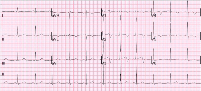
Typically an EKG captures 10 seconds of a cardiac rhythm. The very last line of an EKG is called the rhythm strip.  So, if you count the number of beats in that 10 second strip and multiply that number by 6, you get the heart rate.  In the picture above, that is 11 beats in 10 seconds, which works out to 66bpm. We are all taught how to read an EKG rate differently, but any method that works for you is good.

Rhythm

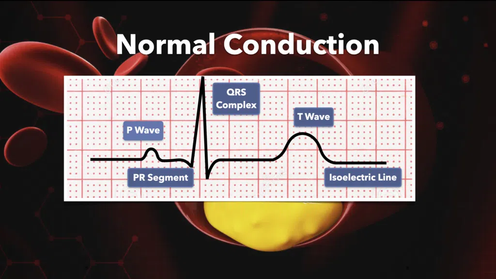
Now that we have the rate, let’s figure out the rhythm.  If we continue to look at that rhythm strip, we will see that the QRS complexes come at a regular interval that steadily marches on across the page.  There is a p-wave for every QRS complex, and a QRS complex for every p-wave.  Also, the QRS complexes all have the same shape.  This defines normal sinus rhythm.  If the rhythm is not regular across the page, in other words some beats came closer together than others, them that would be an irregular rhythm.  Irregular rhythms are not all bad, but you should be able to recognize the bad ones. This is a crucial part of knowing how to read an EKG.

Atrial Fibrillation

Atrial fibrillation is probably the most common, and concerning cause of irregular heart rhythms, and is one that every person who reads EKGs should be familiar with.  In atrial fibrillation, the atria do not beat in a synchronized way, so p-waves are not present on the EKG.  This disorganization causes current to pass through the AV node at different intervals, causing irregular contraction of the ventricles, and therefore irregular intervals between QRS complexes on the EKG.  Atrial fibrillation has an “irregularly irregular” QRS pattern on an EKG.

Premature Contractions

PAC’s and PVC’s are other causes of irregular rhythms on EKGs. “PAC” stands for premature atrial contraction. These are cause by excited or irritated cells in the atria that cause an “early” contraction of the atria. This causes an early QRS complex.  “PVC” stands for premature ventricular contraction. These occur when irritated ventricles cause early depolarization. These are not triggered by the atria.  PVCs do not always translate into ventricular contraction, but they may.  While PACs are usually benign, PVCs can be a sign that the heart is becoming increasingly irritated. This may warrant further investigation by an electrophysiologist.

Random, isolated PVCs are usually a benign condition. Also, PVCs may appear every other beat (bigeminy) or every third beat (trigeminy). However, when PVCs appear right after each other, they are called “couplets” and can be significant.  Three PVCs in a row is the definition of non-sustained ventricular tachycardia.  Episodes of bigeminy and trigemini should get your attention and are benign. However, episodes of non-sustained v-tach need to be investigated.

Intervals

Normal Cardiac Conduction

The intervals we are talking about here is the interval between each small component of the cardiac cycle.  Again, modern EKGs will have all of this information in the “numbers box” at the top of the page.  Thankfully we don’t have to break out the calipers and measure these manually anymore!  The important things to know here are what long intervals mean, and what you need to look for when you find them.

PR Interval

Let’s start with the PR interval.  The PR interval correlates to the time it takes for the atria to depolarize and the electrical current to reach and travel through the AV node.  A normal PR interval is less than 210 milliseconds in duration.  A first degree AV block occurs when the PR interval is longer than 210ms. 1st degree AV block can be a completely benign finding, and is nothing to usually be too concerned over.  However, a new 1st degree AV block may be due to drugs, medications, or electrolyte abnormalities. Knowing how to read an EKG and finding subtle changes he

PR intervals will also change for other kinds of heart blocks as well.  2nd degree heart blocks are classified in two different types.  2nd degree Type 1 heart block has a progressively lengthening PR interval until a QRS complex is dropped.   This does not usually progress into an unstable rhythm and does not require inpatient evaluation.  2nd degree Type 2, however, is a serious finding that does require admission for further evaluation.  It is classified as a steady PR interval with a randomly dropped QRS complex, often times in a set block suck as 2:1.  3rd degree heart block is also known as AV dissociation. Here, the AV node is completely blocked. P waves are no longer in rhythm with the QRS complexes.  This will require hospital admission as well as placement of a pacemaker.

QRS Interval

Next, we will look at the QRS interval.  The QRS interval represents depolarization of the ventricles, and is typically less than 120ms.  Widening of the QRS complex and prolongation of the QRS interval is often associated with pathology. This could be due to a conduction block. A widened QRS, should be investigated if it is a new finding.  Possible causes include drug toxicity, hyperkalemia, hypermagnesemia, WPW, and hypothermia.

QT Interval

Finally, we will look at the QT interval.  The QT interval correlates to the time from the completion of the depolarization of the ventricles to the completion of the repolarization of the ventricles.  This interval has received a lot of press lately as it seems that every medication known to man will cause a prolongation of the QT interval.  A normal QT interval is less than 450ms, but varies by sex, age, and heart rate.  Usually, prolonged QT intervals are not concerning until they get past 500ms.  Often, prolonged QT intervals are caused by medication, which is why we must be careful when prescribing medications that could possibly affect the QT interval, as there can be an additive effect.

Injury Patterns

Now that we have established our patient’s heart rate, heart rhythm, and if they are experiencing any electrical conduction abnormalities, we can start looking at the “exciting” stuff.  When most people look at EKGs, the first thing they think of is “Is my patient having a heart attack”.  EKGs provide so much more information than that, but now is the time to look for patterns of injury.

When evaluating an EKG for injury pattern, the first place we must look is the ST segment.  This portion of the EKG tracing relates to how electricity travels through the heart. This segment Is often elevated above – or depressed below – the isoelectric line. The T-P segment defines the isoelectric state of the cardiac cycle.

ST Elevation and Depression

ST Elevation

Patients who are experiencing an acute MI often have ST elevation in leads associated with the area of the heart affected by the blockage.  To diagnose a ST elevation MI (STEMI), there must be at least 1mm of ST elevation in two contiguous leads on the EKG.  Prior to the acute MI, patients my show ST depression and/or T-wave inversion on EKG when they are experiencing chest pain.  This shows that a portion of the heart is not receiving the amount of oxygen in requires to perform the required tasks.

ST elevation usually occurs in specific leads based on blood vessel anatomy. The location of the injury can identify which part of the heart is injured.  Leads II, III, and aVf are the “inferior” leads and correlate to the right side of the heart and usually the right coronary artery.  This can supplies the low pressure right side of the heart. Leads V1-V3 are the “anterior” or “septal” leads, and often correlate to a lesion in the left anterior descending coronary artery, also known as the “widow maker”.  Leads V4-V6, as well as leads I and aVL, are the “lateral” leads. These are associated with injury to the circumflex artery that leads around the left side of the heart.

Lead aVR – The forgotten lead

This leaves us just with lead aVR.  ST elevation in lead aVR may correlate to a high-grade lesion of the left main coronary artery right as it comes off of the aorta.  This can be controversial, because there isn’t an associated second contiguous lead with aVR, so therefore cannot meet STEMI criteria.  These patients often have the usual acute MI symptoms; chest pain, shortness of breath, diaphoresis, etc.  And if they have ST elevation in aVR, they will likely have ST depression or T-wave inversion (reciprocal changes) in all of the other leads.  Patients who present this way need emergent evaluation by cardiology, but technically are not having a STEMI by definition.

So now you know the basics of how to read an EKG.  This truly is the basics, and there is so much more information that can be gleaned from all of those squiggly lines!  But, that is for another day.

The slides in this presentation are taken from our

Advanced Practice Provider Clinical Skills and Procedure Workshop

Our classic 2-day workshop designed for ALL Advanced Practice Providers. Current identification and management of urgent and also emergent conditions you are most likely to miss. Procedures to match! Learn how to save a life in any practice setting. CERTAINLY A MUST HAVE live course experience. Friday and Saturday.

SAMPLE VIDEO FROM OUR LIVE COURSE:

To learn more about EKGs and other commonly challenging clinical conditions, register for our program!

Leave a Comment

Are you enjoying this article?

If so, you should check out our

Conferences and Workshops

We make hands-on training fun and engaging!

* Highly rated * Add more confidence * See better outcomes *

Live programs are available in:

The Advanced and Difficult Airway Course

The Ultrasound Course

The Clinical Skills & Procedure Workshop

The Clinical Skills & Procedure Workshop + The Airway Course

The Clinical Skills & Procedure Workshop + The Ultrasound Course

Dermatology Overview

Dermatology Essentials

Definition

Cellulitis: infection of dermis and subcutaneous fat

Impetigo: superficial purulent lesions, esp. on face and extremities. Commonly with bullae and/or golden crust

Erysipelas: raised erythematous lesion with clear borders

Folliculitis: hair follicle inflammation. Superficial and limited to the epidermis.

Furunculosis: hair follicle infection that extend to dermis. Multiple = carbuncle

Necrotizing Infection: Deeper SSTI that involve fascial and/or muscle compartments

Etiology

Microbiology

  • Cellulitis: primarily Staph and Strep, incl. MRSA. In immunocomp./diabetics, GNRs also
    • Other etiologies: cat/dog bite P. moltocida; gardening Sporothrix; salt water Vibrio vulnificus; puncture wound → Pseudomonas
  • Impetigo: Strep or Staph
  • Erysipelas: group A Strep usu.
  • Folliculitis/furunculosis: S. aureus, Pseudomonas
  • Necrotizing Infections: Polymicrobial (eg strep and GNRs in Type I, Fournier’s), Group A Strep, S. aureus, Aeromonas hydrophila, Vibrio vulnificus

At risk: athletic teams, military, prison, MSM, communities with MRSA infxn, Diabetic

High risk for more aggressive infection: splenectomy, immunocompromised

Differential Diagnosis

  • Cellulitis
  • Impetigo
  • Erysipelas
  • Folliculitis
  • Furunculosis
  • Necrotizing fasciitis
  • Myonecrosis
  • Calciphylaxis
  • Cutaneous metastasis from neoplasms (especially adenocarcinoma)
  • Graft-versus-host disease (in appropriate population)
  • Sweet syndrome

Patient History

  • Recent trauma to the affected area?
  • Any recent surgeries (hip replacement is risk factor)?
  • Ask about the presence of HIV, diabetes, liver disease, or kidney disease.
  • History of IV drug abuse or subcutaneous injection.
  • Recurrent Cellulitis: Assess for predisposing conditions such as edema, obesity, eczema, venous stasis, and toe web abnormalities.
  • Recurrent Abscesses: Search for local causes such as pilonidal cyst, HS, or foreign body. Consider 5-day decolonization (intranasal mupirosin, daily chlorhexidine). Consider neutrophil disorder if abscesses began in childhood.

Physical Exam

  • Evaluate affected area for erythema, edema, warmth, and pain on palpation.
  • Look for lymphangiitis (erythematous tracks under the skin marking an inflamed lymphatic system), palpate for lymphadenopathy.
  • Assess for evidence of necrotizing infection: systemic toxicity with high temperature, hypotension, disorientation, lethargy, skin discoloration or bullous lesions, anesthesia, firm skin with wooden-hard induration, pain extending beyond cutaneous erythema, pain out of proportion to exam

Work Up

Note: Diagnosis is largely clinical

Laboratory:

  • CBC with diff, ESR/CRP if concern for osteo, CK if concern for necrotizing infection or pyomyositis.
  • Furuncle/pustule can be aspirated for gram stain and culture.
  • For cellulitis, blood cultures are generally low yield, but should be obtained in patients undergoing chemo, neutropenic patients, and those who suffered animal bites.

Imaging:

  • If concern for osteo, xray; consider MRI
  • If concern for necrotizing infection can look for gas in fascial planes on x-ray or CT, but this is highly insensitive

Triage

More serious presentations of skin and soft tissue infections:

  • Toxic shock syndrome: fever, HA, vomiting, myalgias, pharyngitis, diarrhea, diffuse rash with desquamation. Hypotension and shock.
  • Osteomyelitis: infection of bone due to hematogenous seeding or direct spread from overlying focus.
  • Necrotizing fasciitis: infection and necrosis of superficial fascia, subq fat, and deep fascia. Clues: rapidly spreading cellulitis, systemic toxicity (inc TSS), pain out of proportion to exam, bullae formation, gangrene, crepitus. Surgical and medical emergency.
  • Gas gangrene: Clostridial myonecrosis, a fulminant skeletal muscle infection. C. perfringins usually in the setting of trauma; C. septicum in setting of cancer. Surgical and medical emergency.

Treatment

Purulent (furuncle/carbuncle/abscess):

  • Mild: I & D
  • Moderate: I & D, send for culture and sensitives
    • Empiric treatment: Bactrim 1-2 DS tab BID or Doxycycline 100mg BID
    • Defined treatment: MRSA: Bactrim 1-2 DS tab BID, MSSA: Dicloxacillin 250 Q6H or Cephalexin 500 Q6H or Cefadroxil 500mg po q12.
  • Severe: I & D, send for culture and sensitiivies
    • Empiric treatment: Vancomycin or Daptomycin or Linezolid or Ceftaroline
    • Defined treatment: MRSA: similar to empiric, MSSA: Nafcillin or Cefazolin or Clindamycin (if Susceptible)

Nonpurulent (necrotizing infection/cellulitis/erysipelas):

  • Mild: impetigo: topical mupirocin; oral treatment: Penicillin VK or Cephalosporin (eg Cephalexin 500mg PO Q6H) or Dicloxacillin 500mg PO Q6H or Clindamycin 300mg PO Q8H
  • Moderate: IV therapy: penicillin or Cefriaxone or Cefazolin or Clindamycin 300mg PO Q8H or 600mg IV Q8H
  • Severe: emergency surgical evaluation/debridement to rule out necrotizing process
    • Empiric treatment: Vancomycin PLUS Piperacillin/Tazobactam
    • Defined treatment for necrotizing infections:
      • Strep. pyogenes:Penicillin PLUS Clindamycin
      • Vibrio vulnificus:Doxycycline PLUS Ceftazidime
      • Aeromonas hydrophila:Doxycycline PLUS Ciprofloxacin
      • Polymicrobial: Vancomycin PLUS Piperacillin/Tazobactam

Duration of Therapy: 5-7 Days

Treatment Notes:

Erythema may initially worsen with antibiotics 2/2 local bacterial killing.

– For cellulitis, elevation of the affected extremity is essential to treatment.

– For Staph aureus infections (eg suppurative cellulitis) in 2014 at Hopkins susceptibilities were: TMP-SMX 87-88%, Tetracycline 89-91%, and Clindamycin 46-60%.

– For Beta-hemolytic Strep infections (eg non-suppurative cellulitis) all strains are susceptible to penicillin. At Hopkins there are high rates of resistance to TMP-SMX and tetracyclines and variable rates of resistance to Clindamycin.

– If you are concerned for a necrotizing infection, CONSULT SURGERY. Empiric antibiotic treatment with vancomycin (or linezolid) PLUS zosyn (or carbapenem) should be initiated. Clindamycin can be added to inhibit toxin production.

References

  1. Stevens DL, Bisno AL, Chambers HF, et al. Practice guidelines for the diagnosis and management of skin and soft tissue infections: 2014 update by the Infectious Diseases Society of America. Clin Infect Dis. 2014;59(2):e10-52. [PMID:24973422]
  2. Swartz MN. Clinical practice. Cellulitis. N Engl J Med. 2004;350(9):904-12. [PMID:14985488]

Resources

Heart Failure

Cardiology Essentials

Definition

Syndrome characterized by impaired myocardial performance and progressive maladaptive neurohormonal activation of the cardiovascular system leading to circulatory insufficiency to meet the body’s demands.

Systolic heart failure or heart failure with reduced ejection fraction (HFrEF): Clinical diagnosis of heart failure and an EF of less than 50%.

Diastolic heart failure or heart failure with preserved ejection fraction (HFpEF): Clinical signs and symptoms of heart failure with evidence of normal or preserved EF and evidence of abnormal LV diastolic function by Doppler echocardiography or cardiac catheterization

Right heart failure: Majority of cases are a result of left heart failure, although isolated pulmonary diseases can also cause this syndrome.

Etiology

  • Non-ischemic dilated cardiomyopathy (familial or idiopathic)
  • Hypertrophic cardiomyopathy
  • Restrictive cardiomyopathy
  • Cardiomyopathy as a result of fibroelastosis
  • Mitochondrial disease
  • Left ventricular non-compaction
  • Ischemic cardiomyopathy
  • Stress induced cardiomyopathy
  • Valvular obstruction or insufficiency
  • Hypertensive cardiomyopathy
  • Inflammatory (lymphocytic, eosinophilic, giant cell myocarditis)
  • Infectious (Chagas, Lyme disease, HIV, viral, bacterial, or fungal infections)
  • Endocrine disorders (thyroid disease, adrenal insufficiency, pheochromocytoma, acromegaly)
  • Familial storage disease (hemochromatosis, glycogen storage disease, Hurler syndrome, Anderson-Fabry disease)
  • Amyloidosis
  • Connective tissue disease (SLE, polyarteritis nodosa, scleroderma, myositis, sarcoidosis)
  • Muscular dystrophies
  • Neuromuscular disease (Friedreich ataxia, Noonan disease)
  • Toxins (alcohol, anthracyclines, radiation)
  • Tachyarrhythmia

Pathophysiology

Progressive disorder initiated by a form of myocardial injury either sudden (MI or myocarditis) or chronic insults (familial, metabolic, HTN, valve disease, shunting) that result in maladaptive compensatory mechanisms.

These mechanisms include activation of the sympathetic nervous system and activation of the RAS system which overtime lead to pump dysfunction and circulatory collapse.

Differential Diagnosis

Other entities that may look like acute decompensated heart failure:

  • Acute coronary syndrome
  • Interstitial lung disease
  • Pneumonia
  • ARDS
  • Other sources of volume overload such as CKD/ESRD vs cirrhosis, pulmonary hypertension, PE, cardiac tamponade, constrictive or restrictive pericarditis

Patient History

Ask about the signs and symptoms:

  • Worsening dyspnea at rest or exertion?
  • Fatigue?
  • Orthopnea?
  • PND?
  • Weight gain?
  • Increased edema?
  • Lightheadedness?
  • indigestion?
  • Chest heaviness?
  • Fever?
  • Chest pain?
  • Timing of symptom onset?

Ask about triggers of acute decompensation:

  • dietary indiscretion? foods high in Na like lunch meats, chips, canned foods, fast foods?
  • missed medication doses (diuretic)?
  • are they weighing themself daily? adjusting diuretics?
  • any signs or symptoms that an ischemic event has occurred?
  • do they consume alcohol excessively?

Physical Exam

  • Weight gain (if possible look at previous discharge weights)
  • Elevated jugular venous pulsations (Key!), hepatojugular reflux
  • Orthopnea
  • Pulmonary rales
  • Third and/or fourth heart sound
  • Pedal edema
  • Sacral edema in patients who are mostly in bed

Work Up

Laboratory

  • Renal function panel, liver function panel (CMP): Patients who are volume overloaded due to acute decompensated heart failure often have an acute kidney injury and hepatic congestion.
  • Potassium, calcium (CMP), magnesium. May need to check more frequently (e.g. bid) especially if pt will be diuresed.
  • CBC: Anemia is present in up to 40% of patient with heart failure.
  • Consider pro-BNP if volume exam not helpful; compare to prior.
  • If patient is presenting newly with HF and/or etiology is unclear:
    • troponin and lipid profile, especially if HFrEF the pt may need further work up for ischemic disease
    • TSH
    • in the right patient, consider iron studies (hemochromatosis), serum ceruloplasmin (Wilson’s), trypanosoma cruzi IgG (chagas), blood alcohol level or CDT etc.

Imaging

  • ECG, chest x-ray, echocardiography

Other imaging and diagnostic modalities that can be considered based on the patient’s history:

  • Cardiac MR
  • Nuclear imaging
  • Right heart catheterization
  • Left heart catheterization
  • CT angiogram.
  • Endomyocardial biopsy


Triage

Strongly consider step-down or ICU if evidence of decompensation with hypoperfusion (cold and wet):

Altered mental status, Cold extremities, evidence of organ hypoperfusion: increasing lactate or rising creatine, narrow pulse pressures

Risk Stratification

The American College of Cardiology/American Heart Association (ACC/AHA) Heart Failure Classification is a system used to classify heart failure into four stages based on the severity of symptoms and degree of functional impairment.

The four stages of heart failure in the ACC/AHA classification are:

  1. Stage A: At high risk of developing heart failure due to underlying conditions or risk factors such as hypertension, diabetes, or coronary artery disease.

  2. Stage B: Structural heart disease is present, but there are no symptoms of heart failure. This stage includes patients with a history of myocardial infarction (heart attack) or left ventricular remodeling after a cardiac injury.

  3. Stage C: Structural heart disease is present, and there are symptoms of heart failure such as fatigue, shortness of breath, and decreased exercise tolerance. This stage includes patients with past or current symptoms of heart failure who are responding to treatment.

  4. Stage D: Advanced heart failure that is refractory to standard treatments. This stage includes patients with severe symptoms of heart failure at rest, despite maximal medical therapy. Patients in this stage may require advanced interventions such as heart transplant or mechanical circulatory support.

The ACC/AHA Heart Failure Classification is based on a combination of factors, including clinical symptoms, physical examination findings, imaging studies, and laboratory tests. This classification system is useful for guiding treatment decisions and predicting outcomes in patients with heart failure. It can also help clinicians identify patients at high risk for developing heart failure and initiate preventive interventions to improve outcomes.

The New York Heart Association (NYHA) Functional Classification is a system used to classify heart failure into four stages based on the severity of symptoms and degree of functional impairment. The classification system was developed in 1928 and is still widely used today.

New York Heart Association functional classification

The NYHA Functional Classification is based on the patient’s subjective symptoms and limitations related to physical activity. It is often used in clinical practice to assess the severity of heart failure, guide treatment decisions, and predict outcomes. Patients with more severe symptoms are more likely to have poorer outcomes, and may require more aggressive treatment or consideration of advanced interventions, such as heart transplantation or mechanical circulatory support.

It’s important to note that the NYHA Functional Classification is just one aspect of the overall assessment of heart failure and should be used in conjunction with other clinical and diagnostic findings.

The Seattle Heart Failure Model (SHFM) is a clinical prediction model that provides an estimate of the probability of death and other adverse outcomes in patients with heart failure. It was developed to help clinicians make more informed decisions about treatment and to assist in risk stratification of patients with heart failure. The SHFM incorporates a wide range of patient characteristics, including demographics, clinical symptoms, laboratory values, and medication use, to predict the likelihood of various outcomes, such as mortality, hospitalization, and quality of life. The model is based on data from over 11,000 patients with heart failure and has been validated in several independent cohorts. To use the SHFM, a clinician inputs data on the patient’s age, sex, symptoms, medical history, laboratory values, and medication use into a web-based calculator. The model then generates a personalized estimate of the patient’s probability of death and other outcomes at 1 year and 5 years. The SHFM also provides a range of other information, such as the estimated survival time, probability of hospitalization, and predicted quality of life. The SHFM has been shown to have good accuracy in predicting outcomes in patients with heart failure, and it can be useful in guiding treatment decisions and in risk stratification of patients. However, it is important to note that the SHFM is just one tool among many that can be used in the management of heart failure, and it should be used in conjunction with clinical judgment and other diagnostic and prognostic tools.  

The MAGGIC (Meta-Analysis Global Group in Chronic Heart Failure) risk score is a prognostic model that is used to predict mortality in patients with chronic heart failure. It was developed using a large international database of over 39,000 patients with heart failure from 30 different studies.

The MAGGIC risk score takes into account a range of patient characteristics and clinical features that have been shown to be predictive of mortality in heart failure, including age, sex, systolic blood pressure, NYHA functional class, heart rate, serum sodium, serum creatinine, ejection fraction, etiology of heart failure, and use of certain medications such as ACE inhibitors, beta blockers, and diuretics.

The MAGGIC risk score assigns points to each of these variables based on their estimated contribution to mortality risk. The total number of points is then used to estimate the patient’s probability of mortality at 1 year and up to 5 years. The MAGGIC risk score has been shown to have good discrimination and calibration in predicting mortality in patients with heart failure.

The MAGGIC risk score is useful for identifying high-risk patients who may benefit from closer monitoring and more aggressive treatment, as well as for guiding clinical decision-making and communication with patients and families about prognosis. However, it is important to note that the MAGGIC risk score is just one tool among many that can be used in the management of heart failure, and it should be used in conjunction with clinical judgment and other diagnostic and prognostic tools.

CHA2DS2-VASc score: The CHA2DS2-VASc score is a tool used to estimate the risk of stroke in patients with atrial fibrillation. Since atrial fibrillation is a common comorbidity in heart failure, this score can be useful in managing heart failure patients with concurrent atrial fibrillation.

The CHA2DS2-VASc score is a clinical prediction rule that is primarily used to estimate the risk of stroke in patients with non-valvular atrial fibrillation. It is not specifically used in the management of heart failure, but rather in the management of comorbidities that may be present in patients with heart failure.

Patients with heart failure are at an increased risk of developing atrial fibrillation and other cardiovascular diseases, such as stroke, myocardial infarction, and peripheral vascular disease. As such, the CHA2DS2-VASc score can be used in the management of heart failure as a tool to identify patients who are at an increased risk of developing these conditions, and to guide clinical decision-making regarding the use of prophylactic therapies such as anticoagulation.

The CHA2DS2-VASc score takes into account a range of patient characteristics and clinical features that have been shown to be predictive of stroke and other cardiovascular events, including age, sex, history of stroke or transient ischemic attack, hypertension, diabetes, heart failure, and vascular disease. The score assigns points to each variable based on its estimated contribution to the risk of stroke or other cardiovascular events.

While the CHA2DS2-VASc score is not specifically designed for use in heart failure, it is an important tool that can be used to guide clinical decision-making in the management of patients with heart failure and comorbidities. It can help identify patients who may benefit from prophylactic therapies and other interventions aimed at reducing the risk of stroke and other cardiovascular events.


Heart failure with reduced ejection fraction (HFrEF) is a condition where the heart muscle weakens and can’t pump blood effectively. Treatment for HFrEF usually involves a combination of lifestyle changes, medication, and other interventions.

The HFrEF therapy calculator is a tool that can help healthcare professionals determine the most appropriate treatment plan for patients with HFrEF. The calculator takes into account the patient’s age, sex, blood pressure, kidney function, and other factors, and recommends medications and doses that have been shown to be effective in treating HFrEF.

The calculator is based on guidelines developed by the American College of Cardiology, American Heart Association, and Heart Failure Society of America. These guidelines recommend a combination of medications that target different aspects of heart failure, including angiotensin-converting enzyme (ACE) inhibitors, angiotensin receptor blockers (ARBs), beta blockers, and aldosterone antagonists.

The HFrEF therapy calculator takes into account the patient’s current medications and adjusts the recommendations accordingly. It also provides guidance on when to start or stop certain medications, and how to titrate the doses to achieve the maximum benefit while minimizing side effects.

The Renal Risk Score is a tool that helps predict the risk of developing acute kidney injury in patients with heart failure who are undergoing intravenous diuretic therapy.

The renal risk score is a tool that is primarily used to estimate a patient’s risk of developing acute kidney injury (AKI) after undergoing cardiac surgery. However, the risk of AKI is also a concern in patients with heart failure, particularly those who are hospitalized or receiving treatment with certain medications.

In patients with heart failure, the risk of AKI is often related to factors such as low cardiac output, fluid overload, and the use of medications that can affect kidney function. Some of these medications include diuretics, angiotensin-converting enzyme inhibitors (ACE inhibitors), angiotensin receptor blockers (ARBs), and nonsteroidal anti-inflammatory drugs (NSAIDs).

Several studies have looked at the use of the renal risk score in patients with heart failure. One study, published in the journal Circulation Heart Failure in 2014, found that the renal risk score was able to predict the risk of AKI in patients hospitalized with heart failure. The study also found that patients with higher renal risk scores were more likely to require dialysis or have a longer hospital stay.

Another study, published in the Journal of Cardiac Failure in 2018, evaluated the use of the renal risk score in patients with heart failure who were receiving treatment with sacubitril/valsartan, a medication used to treat heart failure with reduced ejection fraction. The study found that the renal risk score was able to predict the risk of AKI in these patients and could be used to guide dosing of the medication to minimize the risk of kidney injury.

Overall, while the renal risk score was developed for use in patients undergoing cardiac surgery, it may also be a useful tool for predicting the risk of AKI in patients with heart failure. By identifying patients at higher risk of AKI, healthcare providers can take steps to minimize the risk of kidney injury and improve outcomes for these patients.

Treatment

Acute Decompensated Heart Failure

IV diuresis:

Determine home regimen and try to give an increased dose. Patients with anasarca DO NOT ABSORB ORAL MEDS. Remember patients who are naïve to diuretics may not require high doses for good urine output. As a rule of thumb, the furosemide dose can be initially calculated at 40 (mg) X serum creatinine. Titration will be performed according to initial response. Common diuretics include furosemide, torsemide, metolazone, and Chlorothiazide. For ESRD patients who no longer make urine, volume removal will be via ultrafiltration and may need to be done more aggressively as tolerated by BP. Be sure to check electrolytes twice a day and aggressively supplement (keep K around 4 and magnesium around 2.4. Check daily weights (standing if possible) and monitor Ins and Outs.

Afterload reduction in systolic heart failure:

If no kidney injury is detected you can consider an ACE-Inhibitor, otherwise hydralazine with/or without nitrates. In more severe cases, one may consider sodium nitroprusside

Inotropy: Usually in severe cases or if effective diuresis is not achieved despite other efforts.

Dobutamine or milrinone

Remember to hold beta blockers in acute decompensated heart failure

Chronic Heart Failure Therapies

Mortality reducing agents:

  • ACE inhibitors/ARBs
    • start in all pt’s with current or prior sx’s of HFrEF unless contraindicated; try ACEi first and then try ARB if not tolerated
    • caution in pts with ↓SBP, renal insufficiency, or ↑serum potassium (>5.0 mEq/L). Angioedema occurs in < 1% of pts with ACE inhibitors.
  • ANRIs (angiotensin receptor–neprilysin inhibitor: valsartan/sacubitril)
    • start in pt’s with NYHA class II-III HFrEF who tolerate an ACE inhibitor or ARB, replacement by an ARNI is recommended to further reduce morbidity and mortality. Harmful if started concomitantly with ACEi/ARB – wait 36 hrs after stopping ACEi/ARB to inititate
  • Beta blockers (metoprolol succinate, bisoprolol, and carvedilol)
    • start in all pt’s with current or prior sx’s of HFrEF unless contraindicated
  • ISDN + Hydralazine
    • clear benifit in African American pt’s with NYHA class III-IV HFrEF
    • likely beneficial for all pt’s with HFrEF, though utility somewhat limited by TID dosing
  • Aldosterone receptor blockers (eplerenone, spironolactone)
    • recommended in patients with NYHA class II–IV HF and who have LVEF of 35% or less

HF Hospitalization Reducing Agents

  • Digoxin
  • Ivabradine (inhibits the If current in the SA node, ↓HR)
    • can use in NYHA class II-III stable chronic HFrEF (LVEF ≤35%) who tolerate maximum BB in NSR with HR of 70 bpm or more at rest[2]

Advanced Therapies

  • Left ventricular assist device (right heart must be able to tolerate this)
  • Heart transplantation

References

  1. Khot UN, Jia G, Moliterno DJ, et al. Prognostic importance of physical examination for heart failure in non-ST-elevation acute coronary syndromes: the enduring value of Killip classification. JAMA. 2003;290(16):2174-81. [PMID:14570953]
  2. Yancy CW, et al: 2016 ACC/AHA/HFSA Focused Update on NewPharmacological Therapy for Heart Failure: An Update of the 2013 ACCF/AHA Guideline for theManagement of Heart Failure, Journal of the American College of Cardiology (2016), doi: 10.1016/j.jacc.2016.05.011.
  3. Griffin BP, Callahan TD, Menon V, et al. Manual of Cardiovascular Medicine. Lippincott Williams & Wilkins. 2013 4th edition; Heart Failure and Transplant 125-159
  4. Yancy CW, Jessup M, Bozkurt B, et al. 2013 ACCF/AHA guideline for the management of heart failure: a report of the American College of Cardiology Foundation/American Heart Association Task Force on Practice Guidelines. J Am Coll Cardiol. 2013;62(16):e147-239. [PMID:23747642]

Resources

Advanced and Difficult Airway Course Content

Airway Anatomy and Assessment

  • Overview of airway anatomy
  • Indications of normal airway
  • Identification of difficult airway characteristics
  • Application of assessment for anticipation

Indications for Airway Management

  • Predictors of airway failure
  • Identification of clinical risk factors
  • When to intervene

Non-Invasive Airway Interventions

  • Progressive Oxygenation
  • Pulmonary Mechanics
  • Measured oxygenation techniques
  • CPAP and BIPAP

Invasive Airway Management

  • Rescue airway interventions
  • Laryngeal mask airway
  • Posterior pharyngeal airway adjuncts
  • Endotracheal intubation

Airway Options

  • Identification and selection of the correct airway device
  • Discussion of equipment options and their uses
  • Preparation for induction and intubation

Intubation Drugs

  • A pharmacology review of induction agents
  • Selection of the best drug combinations for induction
  • Safe decision-making

Induction and Intubation

  • Application of each prior learning topic to provide safe intubation
  • Stepwise, thorough discussion about safe airway induction and intubation
  • Correct techniques for endotracheal tube insertion

Confirming Tube Placement

  • Processes to ensure an endotracheal tube is correctly placed
  • Discussion about reintubation and confirmation
  • Application to difficult airway management

Difficult Airway Equipment

  • Intubating LMA, Lighted Stylet, Light Wand, Video Laryngoscope and more
  • Learn how to select the correct equipment for the situation
  • Double set-up indications

The Difficult Airway 

  • Application of difficult airway algorithms to fit the correct clinical conditions
  • A stepwise process discussion to maximize airway success
  • Indications and procedure for emergent cricothyrotomy

Ultrasound Course Content

Introduction to Ultrasound

  • Ultrasound Physics
  • Probe functions and types
  • Methods of scanning (sliding, rocking, other movements and techniques)
  • Probe Settings (depth, “knobology”)
  • Hand movements and dexterity

Biliary Ultrasound

  • Liver and Gall Bladder
  • Identification of anatomy
  • Abnormal Findings and Diagnostic Criteria
  • Interpretation of findings and Management

DVT Ultrasound

  • Lower extremity venous anatomy and scanning technique
  • Expected normal and abnormal findings
  • Interpretation of doppler and compressibility images
  • Clinical decision-making and follow-up

eFAST Ultrasound

  • Trauma-focused exam 
  • Includes abdomen, bladder, cardiac, and pulmonary imaging
  • Diagnostic criteria and decision-making
  • Includes common and subtle findings

Ocular Ultrasound

  • Technique and probe placement
  • Retinal detachment, foreign bodies, lens dislocation, vitreous hemorrhage, retrobulbar hematoma, and papilledema
  • Next clinical steps

Pelvic Ultrasound

  • Probe placement and scanning technique
  • Uterine, ovarian, and adnexal pathology
  • Next clinical steps

Pulmonary Ultrasound

  • Small groups, team led with hands-on guidance and findings
  • Review and individual practice sessions with instructors
  • Additional ultrasound applications
  • Wrap-Up and Closing

Renal Ultrasound

  • Anatomy and probe placement
  • Ultrasonic anatomy and expected findings
  • Clinical correlation and management

Soft Tissue Ultrasound

  • Probe selection and settings
  • Foreign body, cellulitis, abscess, and cyst identification
  • Next clinical steps

Ultrasound for Vascular Access

  • Anatomy of peripheral and central veins
  • Application of Ultrasound to assist with line placement
  • Visualization of landmarks and expected clinical findings

Abdominal Aorta Ultrasound

  • Anatomy and ultrasound placement
  • Expected normal and abnormal findings
  • Next clinical steps and application

3-Day Clinical Skills & Procedure Workshop + The Airway Course

Day 1 Morning

Airway Anatomy and Assessment

  • How to assess an airway
  • Identification of landmarks
  • Predicting a difficult airway
  • Special scenarios
  • Airway classification and grading

Indications for Airway Management

  • Clinical conditions
  • Respiratory status
  • Anatomy
  • Predictors of airway need
  • Common approaches

Non-Invasive and Invasive Airway Management

  • Escalation of intervention
  • Sequential oxygenation
  • BIPAP
  • CPAP
  • Endotracheal Intubation
  • Airway Adjuncts (LMA, OPA)

Airway Options and Medications

  • Types of airway devices
  • Airway equipment
  • Laryngoscopes
  • Fiberoptic and Video Scopes
  • Induction agents and dosing

Induction and Intubation

  • Procedure organization and setup
  • Stepwise airway protocols
  • Anatomy
  • Endotracheal Intubation
  • Hands-On Procedure Practice

Tube Confirmation and Difficult Airway Management

  • Confirmatory tests
  • Defining an intact airway
  • How to manage a failed airway
  • Difficult airway algorithm and management
  • Fiberoptic laryngoscopy
  • Laryngeal mask airway
  • Video laryngoscope

Day 1 Afternoon

procedure

Difficult Airway Algorithm and Simulation

  • Application of the difficult airway algorithm
  • Simulated patient scenarios
  • Intubation with video laryngoscopy
  • Hands-On airway procedure lab
  • Individual review with instructor
  • Certification Examination

Day 2 Morning

Cardiac

Cardiac Disorders

  • Course Introduction
  • Cardiac Overview
  • EKG Interpretation
  • Acute MI (recognition, management)
  • Common Dysrhythmias
  • Electrolyte Abnormalities and rhythm impacts
  • Condition blocks
  • Bundle Branch Blocks
  • Application to practice
Pulmonary

Pulmonary Disorders

  • Pulmonary Overview
  • Basic Airway Assessment
  • Pneumothorax
  • Asthma Management
  • COPD Management
  • Supplemental Oxygenation
  • Wells Criteria
  • PERC Rule
  • Using D-Dimer
  • Pulmonary Embolism
  • Treatment of Pulmonary Embolism

Introduction to Radiology – Chest and Abdomen

 
  • Overview of Radiograph Interpretation
  • Chest, Shoulder, Clavicle Radiographs
  • Systemic Reading Process
  • Abnormal Radiographs
  • Radiographic Signs of Major Diseases
  • Suggested treatment guidelines based on findings
  • Radiographic Signs of High Impact Injuries
  • Foreign body ingestion, aspiration, and insertion
  • Pediatric foreign body aspiration and management

Day 2 Afternoon

procedure

Procedure Overview

  • Procedural Overview
  • Needle Decompression
  • Chest Tube Insertion
  • Tracheostomy Replacement
  • Shoulder Reduction and Immobilization
  • Upper Extremity Joint Aspiration
  • Trigger Point Injection
  • Nail Trephination
  • Nail Removal
  • Foreign Body and Fish Hook Removal
    Introduction to Suture Techniques
     
suturing

The Suturing Course

  • Suture Clinic and Equipment Introduction
  • Knot Tying
  • Simple Interrupted
  • Simple Running
  • Mattress
  • Subcutaneous/Multiple Layer Closure
  • Staples
  • Skin Adhesive
  • Surgeon’s Knot
  • Buried Knot
  • Billing and Documentation for Sutures
  • Local Injections and Digital Blocks
 
procedure

Procedure Workshop

  • Knee Injection and Aspiration
  • Shoulder Injection
  • Needle Decompression
  • Chest Tube Insertion

Day 3 Morning

Cervical Spine Injuries

  • Long Board and Collar Removal
  • NEXUS Criteria
  • Unstable Fractures
  • Mechanisms of Common Fractures
  • Immobilization
  • Ordering the Correct Studies
  • Correct Consult and Referral
 

Thoracic and Lumbar Spine Injuries

  • Spine form and function
  • Mechanisms of Injury
  • Unstable Fractures
  • Mechanisms of Common Fractures
  • Cauda Equina Syndrome
  • Epidural Abscess
  • Ordering the Correct Studies
  • Correct Consult and Referral
 
Extremity

Upper and Lower Extremity Injuries

  • Speaking Orthopedics
  • Common Patterns of Fractures
  • Common Dislocation and Reduction Techniques
  • Splinting Techniques and Compartment Syndrome
  • Clavicle, Shoulder, Humerus, Elbow, Radius. Ulna. Paired fractures, Wrist and Carpal Bones, Hand
  • Amputations
  • When to Consult Orthopedics
  • When to Consider Transfer/EMS
  • What to send home

Day 3 Afternoon

skin

Skin Conditions Not to Miss

  • Skin and Soft Tissue Conditions
  • Emergent Rash Identification
  • Cellulitis
  • Abscess Incision and Drainage
  • DVT Identification and Decisions Rules
  • Burn Care and Referral Criteria
  • What Not to Send Home
procedure

Procedure Workshop

  • Procedure Clinic
  • Lumbar Puncture
  • Splinting Workshop
  • Intraosseous Access
  • Central Venous Catheter Insertion

3-Day Clinical Skills & Procedure Workshop + The Ultrasound Course

Day 1 Morning

Introduction to Ultrasound

  • Ultrasound Physics
  • Probe functions and types
  • Methods of scanning (sliding, rocking, other movements and techniques)
  • Probe Settings (depth, “knobology”)
  • Hand movements and dexterity
ultrasound

Abdominal Ultrasound

  • Aorta (all views, normal anatomy, pathology)
  • Biliary Quadrant (gallbladder, stones, techniques)
  • Kidney (hydronephrosis, pyelonephritis)
  • Trans-abdominal Pelvis
procedure

Trauma Ultrasound

  • eFAST exam
  • Right upper quadrant imaging
  • Left upper quadrant imaging
  • Bladder Imaging
  • Cardiac Imaging
  • Lung Imaging
procedure

Free Scan with Live Models

  • Small groups, team led with hands-on guidance and findings

Day 1 Afternoon

Specialty Ultrasound

  • Ocular Ultrasound (retinal detachment, foreign bodies)
  • Foreign body imaging
  • Ultrasound-Guided IV and Central access technique
  • Lower Extremity Vascular Ultrasound
procedure

Afternoon Free Scan

  • Small groups, team led with hands-on guidance and findings
  • Review and individual practice sessions with instructors
  • Additional ultrasound applications
  • Wrap-Up and Closing

Day 2 Morning

Cardiac

Cardiac Disorders

  • Course Introduction
  • Cardiac Overview
  • EKG Interpretation
  • Acute MI (recognition, management)
  • Common Dysrhythmias
  • Electrolyte Abnormalities and rhythm impacts
  • Condition blocks
  • Bundle Branch Blocks
  • Application to practice
Pulmonary

Pulmonary Disorders

  • Pulmonary Overview
  • Basic Airway Assessment
  • Pneumothorax
  • Asthma Management
  • COPD Management
  • Supplemental Oxygenation
  • Wells Criteria
  • PERC Rule
  • Using D-Dimer
  • Pulmonary Embolism
  • Treatment of Pulmonary Embolism

Introduction to Radiology – Chest and Abdomen

 
  • Overview of Radiograph Interpretation
  • Chest, Shoulder, Clavicle Radiographs
  • Systemic Reading Process
  • Abnormal Radiographs
  • Radiographic Signs of Major Diseases
  • Suggested treatment guidelines based on findings
  • Radiographic Signs of High Impact Injuries
  • Foreign body ingestion, aspiration, and insertion
  • Pediatric foreign body aspiration and management

Day 2 Afternoon

procedure

Procedure Overview

  • Procedural Overview
  • Needle Decompression
  • Chest Tube Insertion
  • Tracheostomy Replacement
  • Shoulder Reduction and Immobilization
  • Upper Extremity Joint Aspiration
  • Trigger Point Injection
  • Nail Trephination
  • Nail Removal
  • Foreign Body and Fish Hook Removal
    Introduction to Suture Techniques
     
suturing

The Suturing Course

  • Suture Clinic and Equipment Introduction
  • Knot Tying
  • Simple Interrupted
  • Simple Running
  • Mattress
  • Subcutaneous/Multiple Layer Closure
  • Staples
  • Skin Adhesive
  • Surgeon’s Knot
  • Buried Knot
  • Billing and Documentation for Sutures
  • Local Injections and Digital Blocks
 
procedure

Procedure Workshop

  • Knee Injection and Aspiration
  • Shoulder Injection
  • Needle Decompression
  • Chest Tube Insertion

Day 3 Morning

Cervical Spine Injuries

  • Long Board and Collar Removal
  • NEXUS Criteria
  • Unstable Fractures
  • Mechanisms of Common Fractures
  • Immobilization
  • Ordering the Correct Studies
  • Correct Consult and Referral
 

Thoracic and Lumbar Spine Injuries

  • Spine form and function
  • Mechanisms of Injury
  • Unstable Fractures
  • Mechanisms of Common Fractures
  • Cauda Equina Syndrome
  • Epidural Abscess
  • Ordering the Correct Studies
  • Correct Consult and Referral
 
Extremity

Upper and Lower Extremity Injuries

  • Speaking Orthopedics
  • Common Patterns of Fractures
  • Common Dislocation and Reduction Techniques
  • Splinting Techniques and Compartment Syndrome
  • Clavicle, Shoulder, Humerus, Elbow, Radius. Ulna. Paired fractures, Wrist and Carpal Bones, Hand
  • Amputations
  • When to Consult Orthopedics
  • When to Consider Transfer/EMS
  • What to send home

Day 3 Afternoon

skin

Skin Conditions Not to Miss

  • Skin and Soft Tissue Conditions
  • Emergent Rash Identification
  • Cellulitis
  • Abscess Incision and Drainage
  • DVT Identification and Decisions Rules
  • Burn Care and Referral Criteria
  • What Not to Send Home
procedure

Procedure Workshop

  • Procedure Clinic
  • Lumbar Puncture
  • Splinting Workshop
  • Intraosseous Access
  • Central Venous Catheter Insertion

1-Day Advanced and Difficult Airway Course Schedule

Day 1 Morning

Airway Anatomy and Assessment

  • How to assess an airway
  • Identification of landmarks
  • Predicting a difficult airway
  • Special scenarios
  • Airway classification and grading

Indications for Airway Management

  • Clinical conditions
  • Respiratory status
  • Anatomy
  • Predictors of airway need
  • Common approaches

Non-Invasive and Invasive Airway Management

  • Escalation of intervention
  • Sequential oxygenation
  • BIPAP
  • CPAP
  • Endotracheal Intubation
  • Airway Adjuncts (LMA, OPA)

Airway Options and Medications

  • Types of airway devices
  • Airway equipment
  • Laryngoscopes
  • Fiberoptic and Video Scopes
  • Induction agents and dosing

Induction and Intubation

  • Procedure organization and setup
  • Stepwise airway protocols
  • Anatomy
  • Endotracheal Intubation
  • Hands-On Procedure Practice

Tube Confirmation and Difficult Airway Management

  • Confirmatory tests
  • Defining an intact airway
  • How to manage a failed airway
  • Difficult airway algorithm and management
  • Fiberoptic laryngoscopy
  • Laryngeal mask airway
  • Video laryngoscope

Day 1 Afternoon

procedure

Difficult Airway Algorithm and Simulation

  • Application of the difficult airway algorithm
  • Simulated patient scenarios
  • Intubation with video laryngoscopy
  • Hands-On airway procedure lab
  • Individual review with instructor
  • Certification Examination

1-Day Ultrasound Course Schedule

Day 1 Morning

Introduction to Ultrasound

  • Ultrasound Physics
  • Probe functions and types
  • Methods of scanning (sliding, rocking, other movements and techniques)
  • Probe Settings (depth, “knobology”)
  • Hand movements and dexterity
ultrasound

Abdominal Ultrasound

  • Aorta (all views, normal anatomy, pathology)
  • Biliary Quadrant (gallbladder, stones, techniques)
  • Kidney (hydronephrosis, pyelonephritis)
  • Trans-abdominal Pelvis
procedure

Trauma Ultrasound

  • eFAST exam
  • Right upper quadrant imaging
  • Left upper quadrant imaging
  • Bladder Imaging
  • Cardiac Imaging
  • Lung Imaging
procedure

Free Scan with Live Models

  • Small groups, team led with hands-on guidance and findings

Day 1 Afternoon

Specialty Ultrasound

  • Ocular Ultrasound (retinal detachment, foreign bodies)
  • Foreign body imaging
  • Ultrasound-Guided IV and Central access technique
  • Lower Extremity Vascular Ultrasound
procedure

Afternoon Free Scan

  • Small groups, team led with hands-on guidance and findings
  • Review and individual practice sessions with instructors
  • Additional ultrasound applications
  • Wrap-Up and Closing

The APP Clinical Skills and Procedure Workshop Schedule

Day 1 Morning

Cardiac

Cardiac Disorders

  • Course Introduction
  • Cardiac Overview
  • EKG Interpretation
  • Acute MI (recognition, management)
  • Common Dysrhythmias
  • Electrolyte Abnormalities and rhythm impacts
  • Condition blocks
  • Bundle Branch Blocks
  • Application to practice
Pulmonary

Pulmonary Disorders

  • Pulmonary Overview
  • Basic Airway Assessment
  • Pneumothorax
  • Asthma Management
  • COPD Management
  • Supplemental Oxygenation
  • Wells Criteria
  • PERC Rule
  • Using D-Dimer
  • Pulmonary Embolism
  • Treatment of Pulmonary Embolism

Introduction to Radiology – Chest and Abdomen

 
  • Overview of Radiograph Interpretation
  • Chest, Shoulder, Clavicle Radiographs
  • Systemic Reading Process
  • Abnormal Radiographs
  • Radiographic Signs of Major Diseases
  • Suggested treatment guidelines based on findings
  • Radiographic Signs of High Impact Injuries
  • Foreign body ingestion, aspiration, and insertion
  • Pediatric foreign body aspiration and management

Day 1 Afternoon

procedure

Procedure Overview

  • Procedural Overview
  • Needle Decompression
  • Chest Tube Insertion
  • Tracheostomy Replacement
  • Shoulder Reduction and Immobilization
  • Upper Extremity Joint Aspiration
  • Trigger Point Injection
  • Nail Trephination
  • Nail Removal
  • Foreign Body and Fish Hook Removal
    Introduction to Suture Techniques
     
suturing

The Suturing Course

  • Suture Clinic and Equipment Introduction
  • Knot Tying
  • Simple Interrupted
  • Simple Running
  • Mattress
  • Subcutaneous/Multiple Layer Closure
  • Staples
  • Skin Adhesive
  • Surgeon’s Knot
  • Buried Knot
  • Billing and Documentation for Sutures
  • Local Injections and Digital Blocks
 
procedure

Procedure Workshop

  • Knee Injection and Aspiration
  • Shoulder Injection
  • Needle Decompression
  • Chest Tube Insertion

Day 2 Morning

Cervical Spine Injuries

  • Long Board and Collar Removal
  • NEXUS Criteria
  • Unstable Fractures
  • Mechanisms of Common Fractures
  • Immobilization
  • Ordering the Correct Studies
  • Correct Consult and Referral
 

Thoracic and Lumbar Spine Injuries

  • Spine form and function
  • Mechanisms of Injury
  • Unstable Fractures
  • Mechanisms of Common Fractures
  • Cauda Equina Syndrome
  • Epidural Abscess
  • Ordering the Correct Studies
  • Correct Consult and Referral
 
Extremity

Upper and Lower Extremity Injuries

  • Speaking Orthopedics
  • Common Patterns of Fractures
  • Common Dislocation and Reduction Techniques
  • Splinting Techniques and Compartment Syndrome
  • Clavicle, Shoulder, Humerus, Elbow, Radius. Ulna. Paired fractures, Wrist and Carpal Bones, Hand
  • Amputations
  • When to Consult Orthopedics
  • When to Consider Transfer/EMS
  • What to send home

Day 2 Afternoon

skin

Skin Conditions Not to Miss

  • Skin and Soft Tissue Conditions
  • Emergent Rash Identification
  • Cellulitis
  • Abscess Incision and Drainage
  • DVT Identification and Decisions Rules
  • Burn Care and Referral Criteria
  • What Not to Send Home
procedure

Procedure Workshop

  • Procedure Clinic
  • Lumbar Puncture
  • Splinting Workshop
  • Intraosseous Access
  • Central Venous Catheter Insertion

The Clinical Skills & Procedure Workshop + The Ultrasound Course

JULY 4 SAVINGS!

Celebrate Independence day with us!

All of our LIVE Programs through October have been marked down 20%!

PLUS

Use Code FREEDOM23

and take 10% off ALL ONLINE PRODUCTS (Including our 4-day virtual conference)

ACT NOW!

Offer Expires July 7. Excludes hotels and custom group programs.